1WOQ

Crystal Structure of Inorganic Polyphosphate/ATP-Glucomannokinase From Arthrobacter sp. strain KM At 1.8 A Resolution


Experimental Data Snapshot

  • Method: X-RAY DIFFRACTION
  • Resolution: 1.80 Å
  • R-Value Free: 0.223 
  • R-Value Work: 0.193 
  • R-Value Observed: 0.193 

wwPDB Validation   3D Report Full Report


Ligand Structure Quality Assessment 


This is version 1.4 of the entry. See complete history


Literature

Crystal Structure of Bacterial Inorganic Polyphosphate/ATP-glucomannokinase: INSIGHTS INTO KINASE EVOLUTION

Mukai, T.Kawai, S.Mori, S.Mikami, B.Murata, K.

(2004) J Biol Chem 279: 50591-50600

  • DOI: https://doi.org/10.1074/jbc.M408126200
  • Primary Citation of Related Structures:  
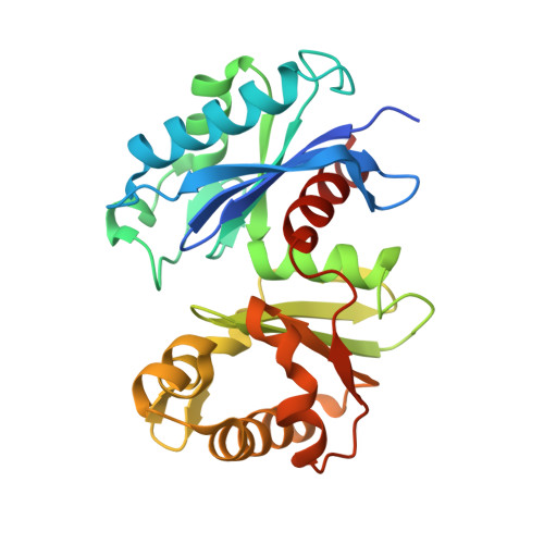
    1WOQ

  • PubMed Abstract: 

    Inorganic polyphosphate (poly(P)) is a biological high energy compound presumed to be an ancient energy carrier preceding ATP. Several poly(P)-dependent kinases that use poly(P) as a phosphoryl donor are known to function in bacteria, but crystal structures of these kinases have not been solved. Here we present the crystal structure of bacterial poly(P)/ATP-glucomannokinase, belonging to Gram-positive bacterial glucokinase, complexed with 1 glucose molecule and 2 phosphate molecules at 1.8 A resolution, being the first among poly(P)-dependent kinases and bacterial glucokinases. The poly(P)/ATP-glucomannokinase structure enabled us to understand the structural relationship of bacterial glucokinase to eucaryotic hexokinase and ADP-glucokinase, which has remained a matter of debate. These comparisons also enabled us to propose putative binding sites for phosphoryl groups for ATP and especially for poly(P) and to obtain insights into the evolution of kinase, particularly from primordial poly(P)-specific to ubiquitous ATP-specific proteins.


  • Organizational Affiliation

    Department of Basic and Applied Molecular Biotechnology, Division of Food and Biological Science, Graduate School of Agriculture, Kyoto University, Uji, Kyoto 611-0011, Japan.


Macromolecules
Find similar proteins by:  (by identity cutoff)  |  3D Structure
Entity ID: 1
MoleculeChains Sequence LengthOrganismDetailsImage
Inorganic polyphosphate/ATP-glucomannokinase
A, B
267Arthrobacter sp. KMMutation(s): 0 
EC: 2.7.1.63
UniProt
Find proteins for Q7WT42 (Arthrobacter sp. (strain KM))
Explore Q7WT42 
Go to UniProtKB:  Q7WT42
Entity Groups  
Sequence Clusters30% Identity50% Identity70% Identity90% Identity95% Identity100% Identity
UniProt GroupQ7WT42
Sequence Annotations
Expand
  • Reference Sequence
Experimental Data & Validation

Experimental Data

  • Method: X-RAY DIFFRACTION
  • Resolution: 1.80 Å
  • R-Value Free: 0.223 
  • R-Value Work: 0.193 
  • R-Value Observed: 0.193 
  • Space Group: P 21 21 21
Unit Cell:
Length ( Å )Angle ( ˚ )
a = 65.214α = 90
b = 82.524β = 90
c = 102.055γ = 90
Software Package:
Software NamePurpose
CNSrefinement
HKL-2000data reduction
SCALEPACKdata scaling
PHASESphasing

Structure Validation

View Full Validation Report


Ligand Structure Quality Assessment 


Entry History 

Deposition Data

Revision History  (Full details and data files)

  • Version 1.0: 2004-09-28
    Type: Initial release
  • Version 1.1: 2008-04-30
    Changes: Version format compliance
  • Version 1.2: 2011-07-13
    Changes: Source and taxonomy, Version format compliance
  • Version 1.3: 2020-07-29
    Type: Remediation
    Reason: Carbohydrate remediation
    Changes: Data collection, Derived calculations, Structure summary
  • Version 1.4: 2024-03-13
    Changes: Data collection, Database references, Structure summary