1WBI

AVR2


Experimental Data Snapshot

  • Method: X-RAY DIFFRACTION
  • Resolution: 1.40 Å
  • R-Value Free: 0.201 
  • R-Value Work: 0.174 
  • R-Value Observed: 0.175 

wwPDB Validation   3D Report Full Report


Ligand Structure Quality Assessment 


This is version 1.3 of the entry. See complete history


Literature

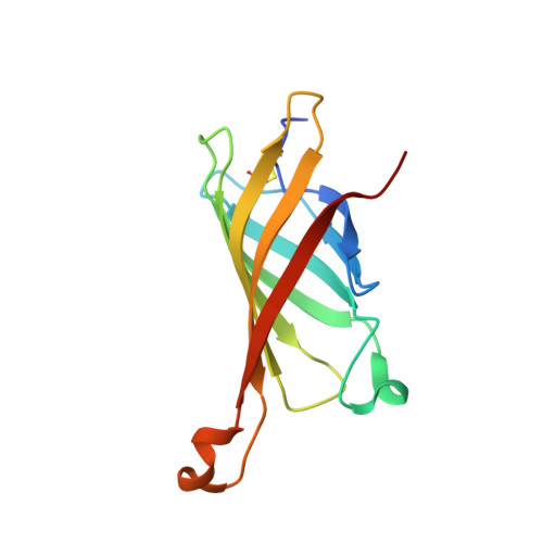
Avidin Related Protein 2 Shows Unique Structural and Functional Features Among the Avidin Protein Family.

Hytonen, V.P.Maatta, J.A.Kidron, H.Halling, K.K.Horha, J.Kulomaa, T.Nyholm, T.K.M.Johnson, M.S.Salminen, T.A.Kulomaa, M.S.Airenne, T.T.

(2005) BMC Biotechnol 5: 28

  • DOI: https://doi.org/10.1186/1472-6750-5-28
  • Primary Citation of Related Structures:  
    1WBI

  • PubMed Abstract: 

    The chicken avidin gene family consists of avidin and several avidin related genes (AVRs). Of these gene products, avidin is the best characterized and is known for its extremely high affinity for D-biotin, a property that is utilized in numerous modern life science applications. Recently, the AVR genes have been expressed as recombinant proteins, which have shown different biotin-binding properties as compared to avidin.


  • Organizational Affiliation

    NanoScience Center, Department of Biological and Environmental Science, University of Jyväskylä, P.O. Box 35 (YAB), FI-40014, Finland. veshyto@bytl.jyu.fi


Macromolecules
Find similar proteins by:  (by identity cutoff)  |  3D Structure
Entity ID: 1
MoleculeChains Sequence LengthOrganismDetailsImage
AVIDIN-RELATED PROTEIN 2
A, B, C, D, E
A, B, C, D, E, F, G, H
129Gallus gallusMutation(s): 0 
UniProt
Find proteins for P56732 (Gallus gallus)
Explore P56732 
Go to UniProtKB:  P56732
Entity Groups  
Sequence Clusters30% Identity50% Identity70% Identity90% Identity95% Identity100% Identity
UniProt GroupP56732
Sequence Annotations
Expand
  • Reference Sequence
Small Molecules
Ligands 3 Unique
IDChains Name / Formula / InChI Key2D Diagram3D Interactions
BTN
Query on BTN

Download Ideal Coordinates CCD File 
EA [auth G]
HA [auth H]
I [auth A]
N [auth B]
R [auth C]
EA [auth G],
HA [auth H],
I [auth A],
N [auth B],
R [auth C],
V [auth D],
W [auth E],
Z [auth F]
BIOTIN
C10 H16 N2 O3 S
YBJHBAHKTGYVGT-ZKWXMUAHSA-N
SO4
Query on SO4

Download Ideal Coordinates CCD File 
CA [auth F]
DA [auth F]
FA [auth G]
GA [auth G]
IA [auth H]
CA [auth F],
DA [auth F],
FA [auth G],
GA [auth G],
IA [auth H],
JA [auth H],
K [auth A],
L [auth A],
M [auth A],
Q [auth B],
T [auth C],
U [auth C],
X [auth E],
Y [auth E]
SULFATE ION
O4 S
QAOWNCQODCNURD-UHFFFAOYSA-L
GOL
Query on GOL

Download Ideal Coordinates CCD File 
AA [auth F]
BA [auth F]
J [auth A]
O [auth B]
P [auth B]
AA [auth F],
BA [auth F],
J [auth A],
O [auth B],
P [auth B],
S [auth C]
GLYCEROL
C3 H8 O3
PEDCQBHIVMGVHV-UHFFFAOYSA-N
Experimental Data & Validation

Experimental Data

  • Method: X-RAY DIFFRACTION
  • Resolution: 1.40 Å
  • R-Value Free: 0.201 
  • R-Value Work: 0.174 
  • R-Value Observed: 0.175 
  • Space Group: P 21 21 21
Unit Cell:
Length ( Å )Angle ( ˚ )
a = 97.669α = 90
b = 99.936β = 90
c = 135.191γ = 90
Software Package:
Software NamePurpose
REFMACrefinement
XDSdata reduction
XDSdata scaling
MOLREPphasing

Structure Validation

View Full Validation Report


Ligand Structure Quality Assessment 


Entry History 

Revision History  (Full details and data files)

  • Version 1.0: 2005-10-12
    Type: Initial release
  • Version 1.1: 2011-05-08
    Changes: Version format compliance
  • Version 1.2: 2011-07-13
    Changes: Version format compliance
  • Version 1.3: 2023-12-13
    Changes: Data collection, Database references, Other, Refinement description