1W9B

S. alba myrosinase in complex with carba-glucotropaeolin


Experimental Data Snapshot

  • Method: X-RAY DIFFRACTION
  • Resolution: 1.70 Å
  • R-Value Free: 0.191 
  • R-Value Work: 0.175 
  • R-Value Observed: 0.175 

wwPDB Validation   3D Report Full Report


Ligand Structure Quality Assessment 


This is version 3.1 of the entry. See complete history


Literature

The glucosinolate-myrosinase system. New insights into enzyme-substrate interactions by use of simplified inhibitors.

Bourderioux, A.Lefoix, M.Gueyrard, D.Tatibouet, A.Cottaz, S.Arzt, S.Burmeister, W.P.Rollin, P.

(2005) Org Biomol Chem 3: 1872-1879

  • DOI: https://doi.org/10.1039/b502990b
  • Primary Citation of Related Structures:  
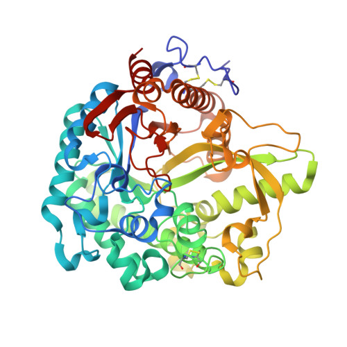
    1W9B

  • PubMed Abstract: 

    Myrosinase, a thioglucoside glucohydrolase, is the only enzyme able to hydrolyse glucosinolates, a unique family of molecules bearing an anomeric O-sulfated thiohydroximate function. Non-hydrolysable myrosinase inhibitors have been devised and studied for their biological interaction. Diverse modifications of the O-sulfate moiety did not result in a significant inhibitory effect, whereas replacing the D-glucopyrano residue by its carba-analogue allowed inhibition to take place. X-Ray experiments carried out after soaking allowed for the first time inclusion of a non-hydrolysable inhibitor inside the enzymatic pocket. Structural tuning of the aglycon part in its pocket is being used as a guide for the development of simplified and more potent inhibitors.


  • Organizational Affiliation

    Institut de Chimie Organique et Analytique (ICOA), UMR 6005, Université d'Orléans, BP 6759, F-45067, Orléans Cedex 2, France.


Macromolecules
Find similar proteins by:  (by identity cutoff)  |  3D Structure
Entity ID: 1
MoleculeChains Sequence LengthOrganismDetailsImage
GLYCOSIDASEA [auth M]501Sinapis albaMutation(s): 0 
EC: 3.2.3.1
UniProt
Find proteins for P29736 (Sinapis alba)
Explore P29736 
Go to UniProtKB:  P29736
Entity Groups  
Sequence Clusters30% Identity50% Identity70% Identity90% Identity95% Identity100% Identity
UniProt GroupP29736
Sequence Annotations
Expand
  • Reference Sequence
Oligosaccharides

Help

Entity ID: 2
MoleculeChains Length2D Diagram Glycosylation3D Interactions
2-acetamido-2-deoxy-beta-D-glucopyranose-(1-4)-2-acetamido-2-deoxy-beta-D-glucopyranoseB [auth A],
E [auth D]
2N-Glycosylation
Glycosylation Resources
GlyTouCan:  G42666HT
GlyCosmos:  G42666HT
GlyGen:  G42666HT
Entity ID: 3
MoleculeChains Length2D Diagram Glycosylation3D Interactions
beta-D-xylopyranose-(1-2)-beta-D-mannopyranose-(1-4)-2-acetamido-2-deoxy-beta-D-glucopyranose-(1-4)-[alpha-L-fucopyranose-(1-3)]2-acetamido-2-deoxy-beta-D-glucopyranoseC [auth B]5N-Glycosylation
Glycosylation Resources
GlyTouCan:  G06206UV
GlyCosmos:  G06206UV
GlyGen:  G06206UV
Entity ID: 4
MoleculeChains Length2D Diagram Glycosylation3D Interactions
beta-D-xylopyranose-(1-2)-[alpha-D-mannopyranose-(1-3)][alpha-D-mannopyranose-(1-6)]beta-D-mannopyranose-(1-4)-2-acetamido-2-deoxy-beta-D-glucopyranose-(1-4)-[alpha-L-fucopyranose-(1-3)]2-acetamido-2-deoxy-beta-D-glucopyranoseD [auth C]7N-Glycosylation
Glycosylation Resources
GlyTouCan:  G26295XE
GlyCosmos:  G26295XE
GlyGen:  G26295XE
Small Molecules
Ligands 5 Unique
IDChains Name / Formula / InChI Key2D Diagram3D Interactions
CGT
Query on CGT

Download Ideal Coordinates CCD File 
L [auth M]CARBA-GLUCOTROPAEOLIN
C15 H21 N O8 S2
LZDZCEOFJWRJIA-GGASBGQWSA-N
NAG
Query on NAG

Download Ideal Coordinates CCD File 
F [auth M]
G [auth M]
H [auth M]
I [auth M]
J [auth M]
F [auth M],
G [auth M],
H [auth M],
I [auth M],
J [auth M],
K [auth M]
2-acetamido-2-deoxy-beta-D-glucopyranose
C8 H15 N O6
OVRNDRQMDRJTHS-FMDGEEDCSA-N
SO4
Query on SO4

Download Ideal Coordinates CCD File 
O [auth M],
P [auth M],
Q [auth M],
R [auth M]
SULFATE ION
O4 S
QAOWNCQODCNURD-UHFFFAOYSA-L
GOL
Query on GOL

Download Ideal Coordinates CCD File 
M,
S [auth M],
T [auth M]
GLYCEROL
C3 H8 O3
PEDCQBHIVMGVHV-UHFFFAOYSA-N
ZN
Query on ZN

Download Ideal Coordinates CCD File 
N [auth M]ZINC ION
Zn
PTFCDOFLOPIGGS-UHFFFAOYSA-N
Binding Affinity Annotations 
IDSourceBinding Affinity
CGT Binding MOAD:  1W9B IC50: 9.90e+5 (nM) from 1 assay(s)
Experimental Data & Validation

Experimental Data

  • Method: X-RAY DIFFRACTION
  • Resolution: 1.70 Å
  • R-Value Free: 0.191 
  • R-Value Work: 0.175 
  • R-Value Observed: 0.175 
  • Space Group: C 2 2 21
Unit Cell:
Length ( Å )Angle ( ˚ )
a = 135.3α = 90
b = 137.2β = 90
c = 80.6γ = 90
Software Package:
Software NamePurpose
CNSrefinement
MOSFLMdata reduction
SCALAdata scaling

Structure Validation

View Full Validation Report


Ligand Structure Quality Assessment 


Entry History 

Deposition Data

Revision History  (Full details and data files)

  • Version 1.0: 2005-05-19
    Type: Initial release
  • Version 1.1: 2011-05-08
    Changes: Version format compliance
  • Version 1.2: 2011-07-13
    Changes: Version format compliance
  • Version 2.0: 2018-12-19
    Changes: Advisory, Atomic model, Data collection, Database references, Derived calculations
  • Version 2.1: 2019-05-08
    Changes: Data collection, Experimental preparation
  • Version 3.0: 2020-07-29
    Type: Remediation
    Reason: Carbohydrate remediation
    Changes: Atomic model, Data collection, Derived calculations, Other, Structure summary
  • Version 3.1: 2023-12-13
    Changes: Data collection, Database references, Derived calculations, Refinement description, Structure summary