


Crystal structure of Putative asparaginyl hydroxylase (2636534) from Bacillus subtilis at 2.60 A resolution
Joint Center for Structural Genomics (JCSG)To be published.
Experimental Data Snapshot
wwPDB Validation 3D Report Full Report

Entity ID: 1 | |||||
|---|---|---|---|---|---|
| Molecule | Chains | Sequence Length | Organism | Details | Image |
| Putative asparaginyl hydroxylase | 342 | Bacillus subtilis | Mutation(s): 6 Gene Names: yxbC | 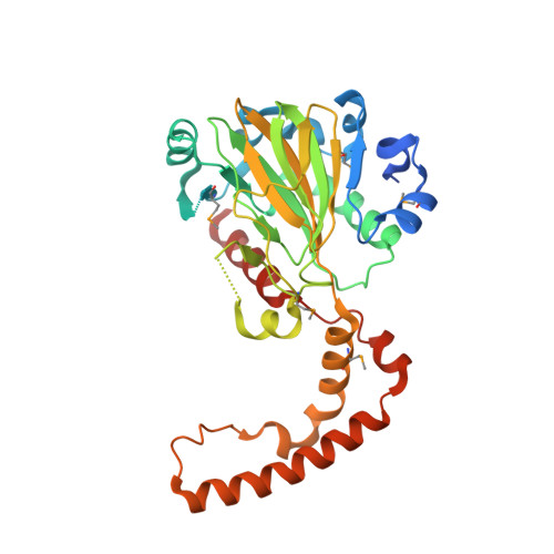 | |
UniProt | |||||
Find proteins for P46327 (Bacillus subtilis (strain 168)) Explore P46327 Go to UniProtKB: P46327 | |||||
Entity Groups | |||||
| Sequence Clusters | 30% Identity50% Identity70% Identity90% Identity95% Identity100% Identity | ||||
| UniProt Group | P46327 | ||||
Sequence AnnotationsExpand | |||||
| |||||
| Ligands 1 Unique | |||||
|---|---|---|---|---|---|
| ID | Chains | Name / Formula / InChI Key | 2D Diagram | 3D Interactions | |
| FE Query on FE | E [auth A], F [auth B], G [auth C], H [auth D] | FE (III) ION Fe VTLYFUHAOXGGBS-UHFFFAOYSA-N |  | ||
| Modified Residues 1 Unique | |||||
|---|---|---|---|---|---|
| ID | Chains | Type | Formula | 2D Diagram | Parent |
| MSE Query on MSE | A, B, C, D | L-PEPTIDE LINKING | C5 H11 N O2 Se |  | MET |
| Length ( Å ) | Angle ( ˚ ) |
|---|---|
| a = 47.3 | α = 90 |
| b = 104.66 | β = 90 |
| c = 286.61 | γ = 90 |
| Software Name | Purpose |
|---|---|
| XDS | data scaling |
| SCALA | data scaling |
| SHELXD | phasing |
| REFMAC | refinement |
| XDS | data reduction |
| CCP4 | data scaling |
| autoSHARP | phasing |
RCSB PDB (citation) is hosted by
RCSB PDB is a member of the

