1UX1

Bacillus subtilis cytidine deaminase with a Cys53His and an Arg56Gln substitution


Experimental Data Snapshot

  • Method: X-RAY DIFFRACTION
  • Resolution: 2.36 Å
  • R-Value Free: 0.245 
  • R-Value Work: 0.213 
  • R-Value Observed: 0.213 

wwPDB Validation   3D Report Full Report


Ligand Structure Quality Assessment 


This is version 1.4 of the entry. See complete history


Literature

Structural, Kinetic, and Mutational Studies of the Zinc Ion Environment in Tetrameric Cytidine Deaminase

Johansson, E.Neuhard, J.Willemoes, M.Larsen, S.

(2004) Biochemistry 43: 6020

  • DOI: https://doi.org/10.1021/bi035893x
  • Primary Citation of Related Structures:  
    1UWZ, 1UX0, 1UX1

  • PubMed Abstract: 

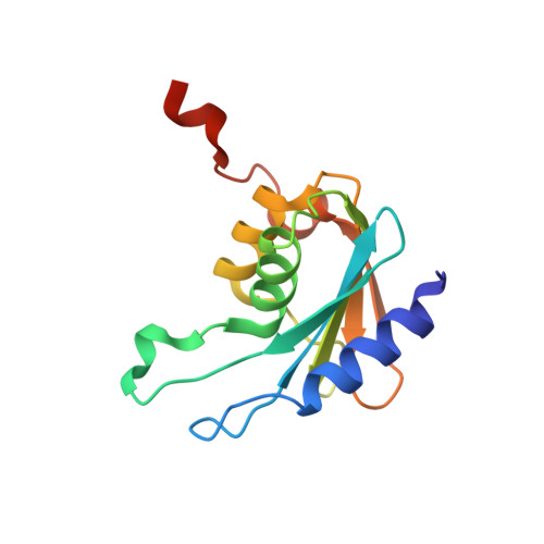
    The zinc-containing cytidine deaminase (CDA, EC 3.5.4.5) is a pyrimidine salvage enzyme catalyzing the hydrolytic deamination of cytidine and 2'-deoxycytidine forming uridine and 2'-deoxyuridine, respectively. Homodimeric CDA (D-CDA) and homotetrameric CDA (T-CDA) both contain one zinc ion per subunit coordinated to the catalytic water molecule. The zinc ligands in D-CDA are one histidine and two cysteine residues, whereas in T-CDA zinc is coordinated to three cysteines. Two of the zinc coordinating cysteines in T-CDA form hydrogen bonds to the conserved residue Arg56, and this residue together with the dipole moments from two alpha-helices partially neutralizes the additional negative charge in the active site, leading to a catalytic activity similar to D-CDA. Arg56 has been substituted by a glutamine (R56Q), the corresponding residue in D-CDA, an alanine (R56A), and an aspartate (R56D). Moreover, one of the zinc-liganding cysteines has been substituted by histidine to mimic D-CDA, alone (C53H) and in combination with R56Q (C53H/R56Q). R56A, R56Q, and C53H/R56Q contain the same amount of zinc as the wild-type enzyme. The zinc-binding capacity of R56D is reduced. Only R56A, R56Q, and C53H/R56Q yielded measurable CDA activity, R56A and R56Q with similar K(m) but decreased V(max) values compared to wild-type enzyme. Because of dissociation into its inactive subunits, it was impossible to determine the kinetic parameters for C53H/R56Q. R56A and C53H/R56Q display increased apparent pK(a) values compared to the wild-type enzyme and R56Q. On the basis of the structures of R56A, R56Q, and C53H/R56Q an explanation is provided of kinetic results and the apparent instability of C53H/R56Q.


  • Organizational Affiliation

    Centre for Crystallographic Studies, Department of Chemistry, University of Copenhagen, Universitetsparken 5, DK-2100 Copenhagen Ø, Denmark. eva@ccs.ki.ku.dk


Macromolecules
Find similar proteins by:  (by identity cutoff)  |  3D Structure
Entity ID: 1
MoleculeChains Sequence LengthOrganismDetailsImage
CYTIDINE DEAMINASE
A, B, C, D
136Bacillus subtilisMutation(s): 2 
EC: 3.5.4.5
UniProt
Find proteins for P19079 (Bacillus subtilis (strain 168))
Explore P19079 
Go to UniProtKB:  P19079
Entity Groups  
Sequence Clusters30% Identity50% Identity70% Identity90% Identity95% Identity100% Identity
UniProt GroupP19079
Sequence Annotations
Expand
  • Reference Sequence
Small Molecules
Ligands 3 Unique
IDChains Name / Formula / InChI Key2D Diagram3D Interactions
THU
Query on THU

Download Ideal Coordinates CCD File 
E [auth A],
H [auth B],
J [auth C],
L [auth D]
TETRAHYDRODEOXYURIDINE
C9 H14 N2 O5
XMJRLEURHMTTRX-FWHJPCMOSA-N
TRS
Query on TRS

Download Ideal Coordinates CCD File 
G [auth A]2-AMINO-2-HYDROXYMETHYL-PROPANE-1,3-DIOL
C4 H12 N O3
LENZDBCJOHFCAS-UHFFFAOYSA-O
ZN
Query on ZN

Download Ideal Coordinates CCD File 
F [auth A],
I [auth B],
K [auth C],
M [auth D]
ZINC ION
Zn
PTFCDOFLOPIGGS-UHFFFAOYSA-N
Experimental Data & Validation

Experimental Data

  • Method: X-RAY DIFFRACTION
  • Resolution: 2.36 Å
  • R-Value Free: 0.245 
  • R-Value Work: 0.213 
  • R-Value Observed: 0.213 
  • Space Group: P 32 2 1
Unit Cell:
Length ( Å )Angle ( ˚ )
a = 62.484α = 90
b = 62.484β = 90
c = 221.604γ = 120
Software Package:
Software NamePurpose
CNSrefinement
DENZOdata reduction
SCALEPACKdata scaling
EPMRphasing

Structure Validation

View Full Validation Report


Ligand Structure Quality Assessment 


Entry History 

Deposition Data

Revision History  (Full details and data files)

  • Version 1.0: 2004-05-20
    Type: Initial release
  • Version 1.1: 2011-05-08
    Changes: Version format compliance
  • Version 1.2: 2011-07-13
    Changes: Version format compliance
  • Version 1.3: 2019-03-06
    Changes: Data collection, Experimental preparation, Other
  • Version 1.4: 2023-12-13
    Changes: Data collection, Database references, Derived calculations, Other, Refinement description