1UU4

X-RAY CRYSTAL STRUCTURE OF THE CATALYTIC DOMAIN OF HUMICOLA GRISEA CEL12A IN COMPLEX WITH CELLOBIOSE


Experimental Data Snapshot

  • Method: X-RAY DIFFRACTION
  • Resolution: 1.49 Å
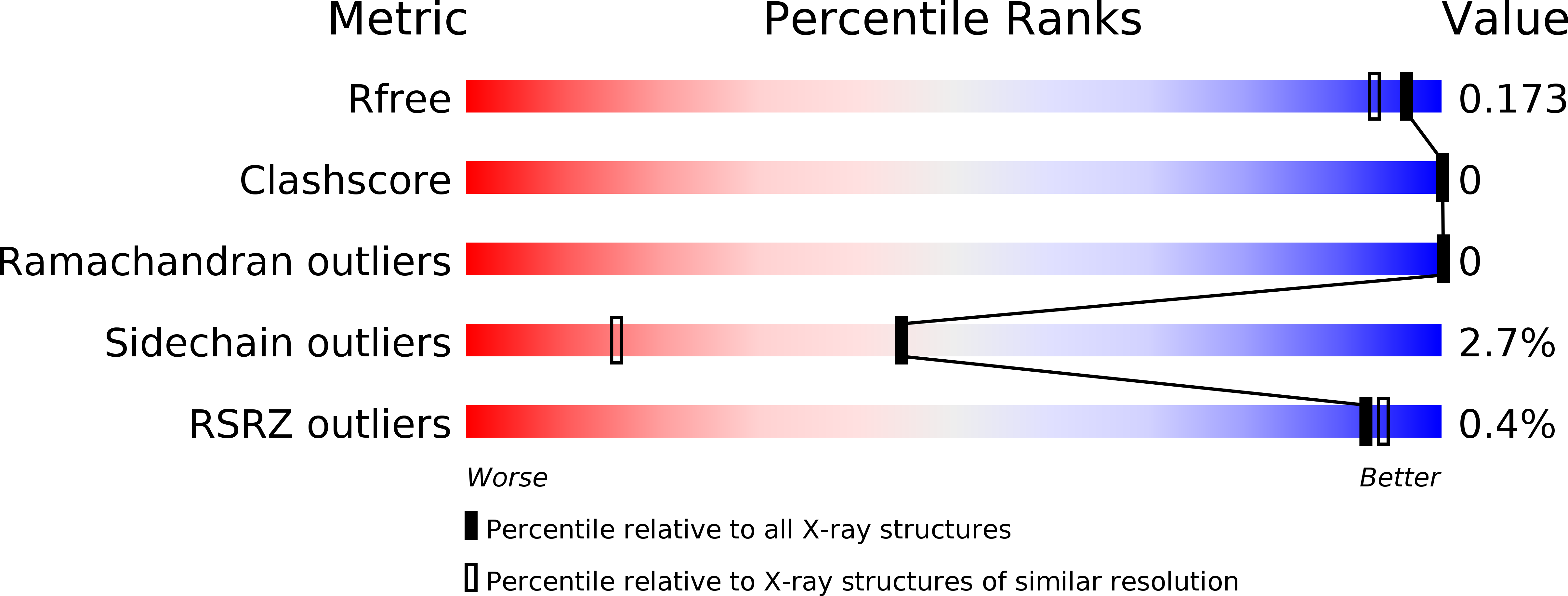
  • R-Value Free: 0.161 
  • R-Value Work: 0.152 
  • R-Value Observed: 0.153 

wwPDB Validation   3D Report Full Report


This is version 3.0 of the entry. See complete history


Literature

Crystal Complex Structures Reveal How Substrate is Bound in the -4 to the +2 Binding Sites of Humicola Grisea Cel12A

Sandgren, M.Berglund, G.I.Shaw, A.Stahlberg, J.Kenne, L.Desmet, T.Mitchinson, C.

(2004) J Mol Biol 342: 1505

  • DOI: https://doi.org/10.1016/j.jmb.2004.07.098
  • Primary Citation of Related Structures:  
    1UU4, 1UU5, 1UU6, 1W2U

  • PubMed Abstract: 

    As part of an ongoing enzyme discovery program to investigate the properties and catalytic mechanism of glycoside hydrolase family 12 (GH 12) endoglucanases, a GH family that contains several cellulases that are of interest in industrial applications, we have solved four new crystal structures of wild-type Humicola grisea Cel12A in complexes formed by soaking with cellobiose, cellotetraose, cellopentaose, and a thio-linked cellotetraose derivative (G2SG2). These complex structures allow mapping of the non-covalent interactions between the enzyme and the glucosyl chain bound in subsites -4 to +2 of the enzyme, and shed light on the mechanism and function of GH 12 cellulases. The unhydrolysed cellopentaose and the G2SG2 cello-oligomers span the active site of the catalytically active H.grisea Cel12A enzyme, with the pyranoside bound in subsite -1 displaying a S31 skew boat conformation. After soaking in cellotetraose, the cello-oligomer that is found bound in site -4 to -1 contains a beta-1,3-linkage between the two cellobiose units in the oligomer, which is believed to have been formed by a transglycosylation reaction that has occurred during the ligand soak of the protein crystals. The close fit of this ligand and the binding sites occupied suggest a novel mixed beta-glucanase activity for this enzyme.


  • Organizational Affiliation

    Department of Cell and Molecular Biology, Uppsala University, Biomedical Center, Box 596, SE-751 24 Uppsala, Sweden. mats@xray.bmc.uu.se


Macromolecules
Find similar proteins by:  (by identity cutoff)  |  3D Structure
Entity ID: 1
MoleculeChains Sequence LengthOrganismDetailsImage
ENDO-BETA-1,4-GLUCANASE224Trichocladium griseumMutation(s): 0 
EC: 3.2.1.4
UniProt
Find proteins for Q8NJY3 (Trichocladium griseum)
Explore Q8NJY3 
Go to UniProtKB:  Q8NJY3
Entity Groups  
Sequence Clusters30% Identity50% Identity70% Identity90% Identity95% Identity100% Identity
UniProt GroupQ8NJY3
Sequence Annotations
Expand
  • Reference Sequence
Oligosaccharides

Help

Entity ID: 2
MoleculeChains Length2D Diagram Glycosylation3D Interactions
beta-D-glucopyranose-(1-4)-beta-D-glucopyranose
B
2N/A
Glycosylation Resources
GlyTouCan:  G84824ZO
GlyCosmos:  G84824ZO
Small Molecules
Ligands 1 Unique
IDChains Name / Formula / InChI Key2D Diagram3D Interactions
PG4
Query on PG4

Download Ideal Coordinates CCD File 
C [auth A]TETRAETHYLENE GLYCOL
C8 H18 O5
UWHCKJMYHZGTIT-UHFFFAOYSA-N
Modified Residues  1 Unique
IDChains TypeFormula2D DiagramParent
PCA
Query on PCA
A
L-PEPTIDE LINKINGC5 H7 N O3GLN
Biologically Interesting Molecules (External Reference) 1 Unique
Experimental Data & Validation

Experimental Data

  • Method: X-RAY DIFFRACTION
  • Resolution: 1.49 Å
  • R-Value Free: 0.161 
  • R-Value Work: 0.152 
  • R-Value Observed: 0.153 
  • Space Group: P 43 21 2
Unit Cell:
Length ( Å )Angle ( ˚ )
a = 49.309α = 90
b = 49.309β = 90
c = 166.15γ = 90
Software Package:
Software NamePurpose
REFMACrefinement
DENZOdata reduction
SCALEPACKdata scaling

Structure Validation

View Full Validation Report


Entry History 

Deposition Data

Revision History  (Full details and data files)

  • Version 1.0: 2004-09-16
    Type: Initial release
  • Version 1.1: 2011-05-08
    Changes: Version format compliance
  • Version 1.2: 2011-07-13
    Changes: Version format compliance
  • Version 2.0: 2020-03-11
    Changes: Advisory, Data collection, Derived calculations, Polymer sequence
  • Version 3.0: 2020-07-29
    Type: Remediation
    Reason: Carbohydrate remediation
    Changes: Atomic model, Data collection, Derived calculations, Structure summary