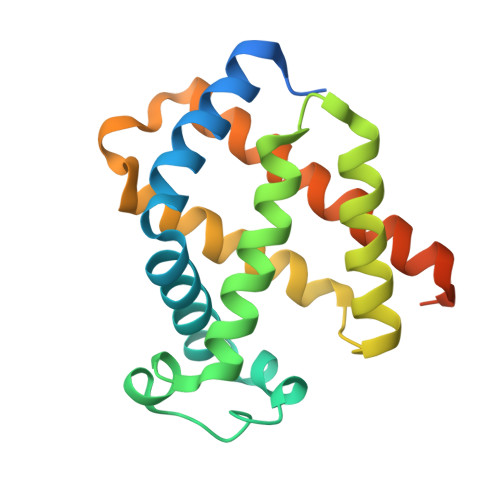
1URY

cytoglobin cavities


Experimental Data Snapshot

  • Method: X-RAY DIFFRACTION
  • Resolution: 2.40 Å
  • R-Value Free: 0.254 
  • R-Value Work: 0.197 
  • R-Value Observed: 0.200 

wwPDB Validation   3D Report Full Report


Ligand Structure Quality Assessment 


This is version 1.2 of the entry. See complete history


Literature

Cytoglobin Cavities

De Sanctis, D.Dewilde, S.Pesce, A.Moens, L.Ascenzi, P.Hankeln, T.Burmester, T.Bolognesi, M.

(2004) Biochem Biophys Res Commun 316: 1217

  • DOI: https://doi.org/10.1016/j.bbrc.2004.03.007
  • Primary Citation of Related Structures:  
    1URY, 1UX9

  • PubMed Abstract: 

    Cytoglobin is the fourth recognized globin type, almost ubiquitously distributed in human tissues; its function is still poorly understood. Cytoglobin displays a core region of about 150 residues, structurally related to hemoglobin and myoglobin, and two extra segments, about 20 residues each, at the N- and C-termini. The core region hosts a large apolar cavity, held to provide a ligand diffusion pathway to/from the heme, and/or ligand temporary docking sites. Here we report the crystal structure (2.4A resolution, R-factor 19.1%) of a human cytoglobin mutant bearing the CysB2(38) --> Ser and CysE9(83) --> Ser substitutions (CYGB*), treated under pressurized xenon. Three Xe atoms bind to the heme distal site region of CYGB* mapping the protein matrix apolar cavity. Despite the conserved globin fold, the cavity found in CYGB* is structured differently from those recognized to play a functional role in myoglobin, neuroglobin, truncated hemoglobins, and Cerebratulus lacteus mini-hemoglobin.


  • Organizational Affiliation

    Department of Physics-INFM and Centre for Excellence in Biomedical Research, University of Genova, Via Dodecaneso 33, Genoa I-16146, Italy.


Macromolecules
Find similar proteins by:  (by identity cutoff)  |  3D Structure
Entity ID: 1
MoleculeChains Sequence LengthOrganismDetailsImage
CYTOGLOBIN
A, B
190Homo sapiensMutation(s): 2 
UniProt & NIH Common Fund Data Resources
Find proteins for Q8WWM9 (Homo sapiens)
Explore Q8WWM9 
Go to UniProtKB:  Q8WWM9
PHAROS:  Q8WWM9
GTEx:  ENSG00000161544 
Entity Groups  
Sequence Clusters30% Identity50% Identity70% Identity90% Identity95% Identity100% Identity
UniProt GroupQ8WWM9
Sequence Annotations
Expand
  • Reference Sequence
Small Molecules
Ligands 3 Unique
IDChains Name / Formula / InChI Key2D Diagram3D Interactions
HEM
Query on HEM

Download Ideal Coordinates CCD File 
C [auth A],
I [auth B]
PROTOPORPHYRIN IX CONTAINING FE
C34 H32 Fe N4 O4
KABFMIBPWCXCRK-RGGAHWMASA-L
FC6
Query on FC6

Download Ideal Coordinates CCD File 
G [auth A],
M [auth B]
HEXACYANOFERRATE(3-)
C6 Fe N6
HCMVSLMENOCDCK-UHFFFAOYSA-N
XE
Query on XE

Download Ideal Coordinates CCD File 
D [auth A]
E [auth A]
F [auth A]
H [auth A]
J [auth B]
D [auth A],
E [auth A],
F [auth A],
H [auth A],
J [auth B],
K [auth B],
L [auth B]
XENON
Xe
FHNFHKCVQCLJFQ-UHFFFAOYSA-N
Experimental Data & Validation

Experimental Data

  • Method: X-RAY DIFFRACTION
  • Resolution: 2.40 Å
  • R-Value Free: 0.254 
  • R-Value Work: 0.197 
  • R-Value Observed: 0.200 
  • Space Group: P 21 21 21
Unit Cell:
Length ( Å )Angle ( ˚ )
a = 47.848α = 90
b = 70.524β = 90
c = 98.728γ = 90
Software Package:
Software NamePurpose
REFMACrefinement
MOSFLMdata reduction
SCALAdata scaling

Structure Validation

View Full Validation Report


Ligand Structure Quality Assessment 


Entry History 

Deposition Data

Revision History  (Full details and data files)

  • Version 1.0: 2004-12-15
    Type: Initial release
  • Version 1.1: 2011-05-08
    Changes: Version format compliance
  • Version 1.2: 2011-07-13
    Changes: Version format compliance