1UMV

Crystal structure of an acidic, non-myotoxic phospholipase A2 from the venom of Bothrops jararacussu


Experimental Data Snapshot

  • Method: X-RAY DIFFRACTION
  • Resolution: 1.79 Å
  • R-Value Free: 0.229 
  • R-Value Work: 0.161 
  • R-Value Observed: 0.164 

wwPDB Validation   3D Report Full Report


This is version 1.3 of the entry. See complete history


Literature

Crystal Structure of an Acidic Platelet Aggregation Inhibitor and Hypotensive Phospholipase A(2) in the Monomeric and Dimeric States: Insights Into its Oligomeric State

Magro, A.J.Murakami, M.T.Marcussi, S.Soares, A.M.Arni, R.K.Fontes, M.R.

(2004) Biochem Biophys Res Commun 323: 24

  • DOI: https://doi.org/10.1016/j.bbrc.2004.08.046
  • Primary Citation of Related Structures:  
    1U73, 1UMV

  • PubMed Abstract: 

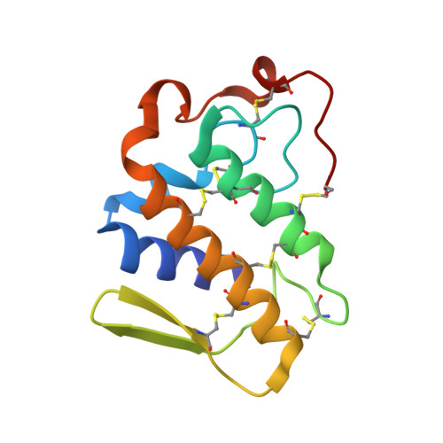
    Phospholipases A2 belong to the superfamily of proteins which hydrolyzes the sn-2 acyl groups of membrane phospholipids to release arachidonic acid and lysophospholipids. An acidic phospholipase A2 isolated from Bothrops jararacussu snake venom presents a high catalytic, platelet aggregation inhibition and hypotensive activities. This protein was crystallized in two oligomeric states: monomeric and dimeric. The crystal structures were solved at 1.79 and 1.90 angstroms resolution, respectively, for the two states. It was identified a Na+ ion at the center of Ca2+-binding site of the monomeric form. A novel dimeric conformation with the active sites exposed to the solvent was observed. Conformational states of the molecule may be due to the physicochemical conditions used in the crystallization experiments. We suggest dimeric state is one found in vivo.


  • Organizational Affiliation

    Departamento de Física e Biofísica, Instituto de Biociências, UNESP, Botucatu-SP, Brazil.


Macromolecules
Find similar proteins by:  (by identity cutoff)  |  3D Structure
Entity ID: 1
MoleculeChains Sequence LengthOrganismDetailsImage
HYPOTENSIVE PHOSPHOLIPASE A2A [auth X]122Bothrops jararacussuMutation(s): 0 
Membrane Entity: Yes 
UniProt
Find proteins for Q8AXY1 (Bothrops jararacussu)
Explore Q8AXY1 
Go to UniProtKB:  Q8AXY1
Entity Groups  
Sequence Clusters30% Identity50% Identity70% Identity90% Identity95% Identity100% Identity
UniProt GroupQ8AXY1
Sequence Annotations
Expand
  • Reference Sequence
Small Molecules
Ligands 1 Unique
IDChains Name / Formula / InChI Key2D Diagram3D Interactions
CA
Query on CA

Download Ideal Coordinates CCD File 
B [auth X]CALCIUM ION
Ca
BHPQYMZQTOCNFJ-UHFFFAOYSA-N
Experimental Data & Validation

Experimental Data

  • Method: X-RAY DIFFRACTION
  • Resolution: 1.79 Å
  • R-Value Free: 0.229 
  • R-Value Work: 0.161 
  • R-Value Observed: 0.164 
  • Space Group: C 2 2 21
Unit Cell:
Length ( Å )Angle ( ˚ )
a = 39.99α = 90
b = 53.99β = 90
c = 90.46γ = 90
Software Package:
Software NamePurpose
REFMACrefinement
DENZOdata reduction
SCALEPACKdata scaling
AMoREphasing

Structure Validation

View Full Validation Report


Entry History 

Deposition Data

Revision History  (Full details and data files)

  • Version 1.0: 2003-09-18
    Type: Initial release
  • Version 1.1: 2011-05-08
    Changes: Version format compliance
  • Version 1.2: 2011-07-13
    Changes: Version format compliance
  • Version 1.3: 2023-12-13
    Changes: Data collection, Database references, Derived calculations, Refinement description