1UCW

COMPLEX OF TRANSALDOLASE WITH THE REDUCED SCHIFF-BASE INTERMEDIATE


Experimental Data Snapshot

  • Method: X-RAY DIFFRACTION
  • Resolution: 2.20 Å
  • R-Value Free: 0.244 
  • R-Value Work: 0.204 
  • R-Value Observed: 0.204 

wwPDB Validation   3D Report Full Report


This is version 1.2 of the entry. See complete history


Literature

Crystal structure of the reduced Schiff-base intermediate complex of transaldolase B from Escherichia coli: mechanistic implications for class I aldolases.

Jia, J.Schorken, U.Lindqvist, Y.Sprenger, G.A.Schneider, G.

(1997) Protein Sci 6: 119-124

  • DOI: https://doi.org/10.1002/pro.5560060113
  • Primary Citation of Related Structures:  
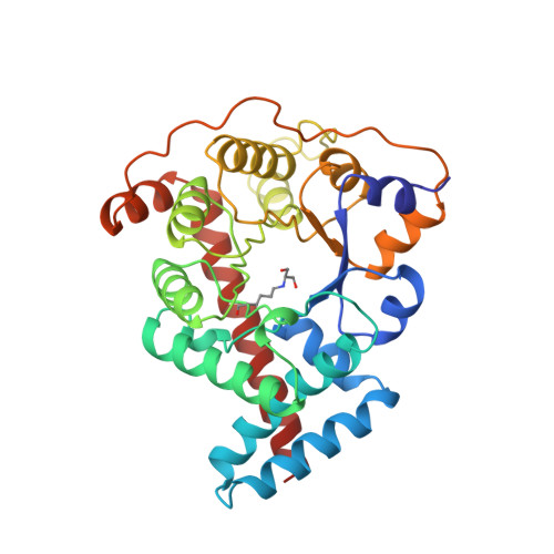
    1UCW

  • PubMed Abstract: 

    Transaldolase catalyzes transfer of a dihydroxyacetone moiety from a ketose donor to an aldose acceptor. During catalysis, a Schiff-base intermediate between dihydroxyacetone and the epsilon-amino group of a lysine residue at the active site of the enzyme is formed. This Schiff-base intermediate has been trapped by reduction with potassium borohydride, and the crystal structure of this complex has been determined at 2.2 A resolution. The overall structures of the complex and the native enzyme are very similar; formation of the intermediate induces no large conformational changes. The dihydroxyacetone moiety is covalently linked to the side chain of Lys 132 at the active site of the enzyme. The Cl hydroxyl group of the dihydroxyacetone moiety forms hydrogen bonds to the side chains of residues Asn 154 and Ser 176. The C3 hydroxyl group interacts with the side chain of Asp 17 and Asn 35. Based on the crystal structure of this complex a reaction mechanism for transaldolase is proposed.


  • Organizational Affiliation

    Department of Medical Biochemistry and Biophysics, Karolinska Institute, Stockholm, Sweden.


Macromolecules
Find similar proteins by:  (by identity cutoff)  |  3D Structure
Entity ID: 1
MoleculeChains Sequence LengthOrganismDetailsImage
TRANSALDOLASE
A, B
317Escherichia coliMutation(s): 1 
EC: 2.2.1.2
UniProt
Find proteins for P0A870 (Escherichia coli (strain K12))
Explore P0A870 
Go to UniProtKB:  P0A870
Entity Groups  
Sequence Clusters30% Identity50% Identity70% Identity90% Identity95% Identity100% Identity
UniProt GroupP0A870
Sequence Annotations
Expand
  • Reference Sequence
Small Molecules
Modified Residues  1 Unique
IDChains TypeFormula2D DiagramParent
LLY
Query on LLY
A, B
L-PEPTIDE LINKINGC9 H16 N2 O6LYS
Experimental Data & Validation

Experimental Data

  • Method: X-RAY DIFFRACTION
  • Resolution: 2.20 Å
  • R-Value Free: 0.244 
  • R-Value Work: 0.204 
  • R-Value Observed: 0.204 
  • Space Group: P 21 21 21
Unit Cell:
Length ( Å )Angle ( ˚ )
a = 69.2α = 90
b = 91.9β = 90
c = 130.9γ = 90
Software Package:
Software NamePurpose
X-PLORmodel building
X-PLORrefinement
DENZOdata reduction
X-PLORphasing

Structure Validation

View Full Validation Report


Entry History 

Deposition Data

Revision History  (Full details and data files)

  • Version 1.0: 1997-07-07
    Type: Initial release
  • Version 1.1: 2008-03-24
    Changes: Version format compliance
  • Version 1.2: 2011-07-13
    Changes: Version format compliance