1U22

A. thaliana cobalamine independent methionine synthase


Experimental Data Snapshot

  • Method: X-RAY DIFFRACTION
  • Resolution: 2.65 Å
  • R-Value Free: 0.270 
  • R-Value Work: 0.200 
  • R-Value Observed: 0.203 

wwPDB Validation   3D Report Full Report


Ligand Structure Quality Assessment 


This is version 1.5 of the entry. See complete history


Literature

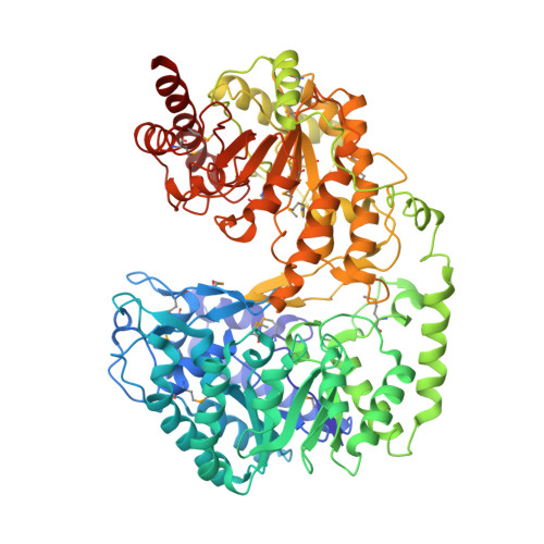
Crystal structures of cobalamin-independent methionine synthase complexed with zinc, homocysteine, and methyltetrahydrofolate

Ferrer, J.-L.Ravanel, S.Robert, M.Dumas, R.

(2004) J Biol Chem 279: 44235-44238

  • DOI: https://doi.org/10.1074/jbc.C400325200
  • Primary Citation of Related Structures:  
    1U1H, 1U1J, 1U1U, 1U22

  • PubMed Abstract: 

    Cobalamin-independent methionine synthase (MetE) catalyzes the synthesis of methionine by a direct transfer of the methyl group of N5-methyltetrahydrofolate (CH3-H2PteGlun) to the sulfur atom of homocysteine (Hcy). We report here the first crystal structure of this metalloenzyme under different forms, free or complexed with the Hcy and folate substrates. The Arabidopsis thaliana MetE (AtMetE) crystals reveal a monomeric structure built by two (betaalpha)8 barrels making a deep groove at their interface. The active site is located at the surface of the C-terminal domain, facing the large interdomain cleft. Inside the active site, His647, Cys649, and Cys733 are involved in zinc coordination, whereas Asp605, Ile437, and Ser439 interact with Hcy. Opposite the zinc/Hcy binding site, a cationic loop (residues 507-529) belonging to the C-terminal domain anchors the first glutamyl residue of CH3-H4PteGlu5. The pterin moiety of CH3-H4PteGlu5 is stacked with Trp567, enabling the N5-methyl group to protrude in the direction of the zinc atom. These data suggest a structural role of the N-terminal domain of AtMetE in the stabilization of loop 507-529 and in the interaction with the poly-glutamate chain of CH3-H4PteGlun. Comparison of AtMetE structures reveals that the addition of Hcy does not lead to a direct coordination of the sulfur atom with zinc but to a reorganization of the zinc binding site with a stronger coordination to Cys649, Cys733, and a water molecule.


  • Organizational Affiliation

    Laboratoire de Cristallogenèse et Cristallographie des Protéines, Institut de Biologie Structurale J.-P. Ebel, 41 rue Jules Horowitz, 38027 Grenoble 1, France. jean-luc.ferrer@ibs.fr


Macromolecules
Find similar proteins by:  (by identity cutoff)  |  3D Structure
Entity ID: 1
MoleculeChains Sequence LengthOrganismDetailsImage
5-methyltetrahydropteroyltriglutamate--homocysteine methyltransferase765Arabidopsis thalianaMutation(s): 19 
Gene Names: CIMS
EC: 2.1.1.14
UniProt
Find proteins for O50008 (Arabidopsis thaliana)
Explore O50008 
Go to UniProtKB:  O50008
Entity Groups  
Sequence Clusters30% Identity50% Identity70% Identity90% Identity95% Identity100% Identity
UniProt GroupO50008
Sequence Annotations
Expand
  • Reference Sequence
Small Molecules
Ligands 4 Unique
IDChains Name / Formula / InChI Key2D Diagram3D Interactions
THG
Query on THG

Download Ideal Coordinates CCD File 
I [auth A](6S)-5,6,7,8-TETRAHYDROFOLATE
C19 H23 N7 O6
MSTNYGQPCMXVAQ-RYUDHWBXSA-N
HCS
Query on HCS

Download Ideal Coordinates CCD File 
H [auth A]2-AMINO-4-MERCAPTO-BUTYRIC ACID
C4 H9 N O2 S
FFFHZYDWPBMWHY-VKHMYHEASA-N
SO4
Query on SO4

Download Ideal Coordinates CCD File 
D [auth A],
E [auth A],
F [auth A],
G [auth A]
SULFATE ION
O4 S
QAOWNCQODCNURD-UHFFFAOYSA-L
ZN
Query on ZN

Download Ideal Coordinates CCD File 
B [auth A],
C [auth A]
ZINC ION
Zn
PTFCDOFLOPIGGS-UHFFFAOYSA-N
Modified Residues  1 Unique
IDChains TypeFormula2D DiagramParent
MSE
Query on MSE
A
L-PEPTIDE LINKINGC5 H11 N O2 SeMET
Experimental Data & Validation

Experimental Data

  • Method: X-RAY DIFFRACTION
  • Resolution: 2.65 Å
  • R-Value Free: 0.270 
  • R-Value Work: 0.200 
  • R-Value Observed: 0.203 
  • Space Group: P 65
Unit Cell:
Length ( Å )Angle ( ˚ )
a = 122.423α = 90
b = 122.423β = 90
c = 131.365γ = 120
Software Package:
Software NamePurpose
XDSdata scaling
XSCALEdata scaling
CNSrefinement
XDSdata reduction
CNSphasing

Structure Validation

View Full Validation Report


Ligand Structure Quality Assessment 


Entry History 

Deposition Data

Revision History  (Full details and data files)

  • Version 1.0: 2004-09-07
    Type: Initial release
  • Version 1.1: 2007-10-16
    Changes: Version format compliance
  • Version 1.2: 2011-07-13
    Changes: Version format compliance
  • Version 1.3: 2023-08-23
    Changes: Data collection, Database references, Derived calculations, Refinement description
  • Version 1.4: 2023-11-15
    Changes: Data collection
  • Version 1.5: 2024-04-03
    Changes: Structure summary