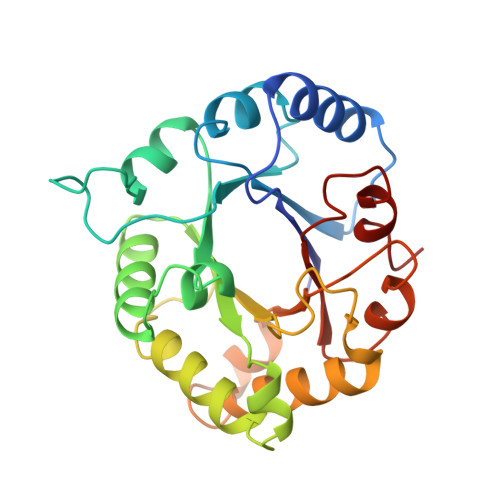
The structural basis for pseudoreversion of the E165D lesion by the secondary S96P mutation in triosephosphate isomerase depends on the positions of active site water molecules.
Komives, E.A., Lougheed, J.C., Liu, K., Sugio, S., Zhang, Z., Petsko, G.A., Ringe, D.(1995) Biochemistry 34: 13612-13621
- PubMed: 7577950
- DOI: https://doi.org/10.1021/bi00041a041
- Primary Citation of Related Structures:
1TPB, 1TPC - PubMed Abstract:
The structural basis for the improvement in catalytic efficiency of the mutant E165D chicken triosephosphate isomerase by the secondary mutation, S96P, has been analyzed using a combination of X-ray crystallography and Fourier transform infrared spectroscopy. All X-ray structures were of the complex of phosphoglycolohydroxamate (PGH), an intermediate analog, with the isomerase, and each was solved to a resolution of 1.9 A. Comparison of the structure of the double mutant, E165D.S96P, with that of the single mutant, E165D, as well as with the wild-type isomerase shows only insignificant differences in the positions of the side chains in all of the mutants when compared with the wild-type isomerase, except that in both the E165D and E165D.S96P mutants, the aspartate side chain was approximately 0.7 A further away from the substrate analog than the glutamate side chain. Significant differences were observed in the crystal structure of the E165D.S96P double mutant in the positions of ordered water molecules bound at the active site. The loss of two water molecules located near the side chain at position 165 was observed in isomerases containing the S96P mutation. The resulting increase in hydrophobicity of the pocket probably causes an increase in the pKa of the catalytic base, D165, thereby improving its basicity. A new ordered water molecule was observed underneath the bound PGH in the E165D.S96P structure, which likely decreases the pKa's of the substrate protons, thereby increasing their acidity. An enzyme derived carbonyl stretch at 1746 cm-1 that is only observed in the IR spectrum of the E165D.S96P double mutant isomerase with bound substrates has been assigned to a stable ground state protonated D165-enediol(ate) intermediate complex. Thus, the gain in activity resulting from the S96P second site change probably results from a combination of improving the basicity of the enzyme, improving the acidity of the substrate protons, and stabilization of a reaction intermediate. All three of these effects seem to be caused by changes in bound water molecules.
Organizational Affiliation:
Department of Chemistry and Biochemistry, University of California, San Diego, La Jolla 92093-0601, USA.