Crystal structure of Escherichia coli thioredoxin reductase refined at 2 A resolution. Implications for a large conformational change during catalysis.
Waksman, G., Krishna, T.S.R., Williams Jr., C.H., Kuriyan, J.(1994) J Mol Biol 236: 800-816
- PubMed: 8114095
- Primary Citation of Related Structures:
1TDE, 1TDF - PubMed Abstract:
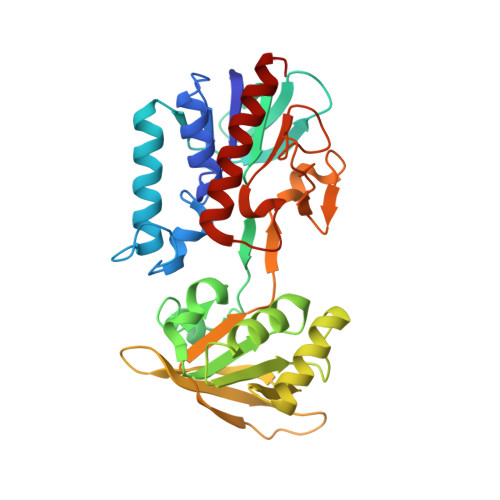
The crystal structures of three forms of Escherichia coli thioredoxin reductase have been refined: the oxidized form of the wild-type enzyme at 2.1 A resolution, a variant containing a cysteine to serine mutation at the active site (Cys138Ser) at 2.0 A resolution, and a complex of this variant with nicotinamide adenine dinucleotide phosphate (NADP+) at 2.3 A resolution. The enzyme mechanism involves the transfer of reducing equivalents from reduced nicotinamide adenine dinucleotide phosphate (NADPH) to a disulfide bond in the enzyme, via a flavin adenine dinucleotide (FAD). Thioredoxin reductase contains FAD and NADPH binding domains that are structurally similar to the corresponding domains of the related enzyme glutathione reductase. The relative orientation of these domains is, however, very different in the two enzymes: when the FAD domains of thioredoxin and glutathione reductases are superimposed, the NADPH domain of one is rotated by 66 degrees with respect to the other. The observed binding mode of NADP+ in thioredoxin reductase is non-productive in that the nicotinamide ring is more than 17 A from the flavin ring system. While in glutathione reductase the redox active disulfide is located in the FAD domain, in thioredoxin reductase it is in the NADPH domain and is part of a four-residue sequence (Cys-Ala-Thr-Cys) that is close in structure to the corresponding region of thioredoxin (Cys-Gly-Pro-Cys), with a root-mean-square deviation of 0.22 A for atoms in the disulfide bonded ring. There are no significant conformational differences between the structure of the wild-type enzyme and that of the Cys138Ser mutant, except that a disulfide bond is not present in the latter. The disulfide bond is positioned productively in this conformation of the enzyme, i.e. it stacks against the flavin ring system in a position that would facilitate its reduction by the flavin. However, the cysteine residues are relatively inaccessible for interaction with the substrate, thioredoxin. These results suggest that thioredoxin reductase must undergo conformational changes during enzyme catalysis. All three structures reported here are for the same conformation of the enzyme and no direct evidence is available as yet for such conformational changes. The simplest possibility is that the NADPH domain rotates between the conformation observed here and an orientation similar to that seen in glutathione reductase. This would alternately place the nicotinamide ring and the disulfide bond near the flavin ring, and expose the cysteine residues for reaction with thioredoxin in the hypothetical conformation.(ABSTRACT TRUNCATED AT 400 WORDS)
Organizational Affiliation:
Laboratory of Molecular Biophysics, Rockefeller University, New York, NY 10021.