Crystallographic studies of V44 mutants of Clostridium pasteurianum rubredoxin: Effects of side-chain size on reduction potential.
Park, I.Y., Eidsness, M.K., Lin, I.J., Gebel, E.B., Youn, B., Harley, J.L., Machonkin, T.E., Frederick, R.O., Markley, J.L., Smith, E.T., Ichiye, T., Kang, C.(2004) Proteins 57: 618-618
- PubMed: 15382226
- DOI: https://doi.org/10.1002/prot.20243
- Primary Citation of Related Structures:
1T9O, 1T9P, 1T9Q - PubMed Abstract:
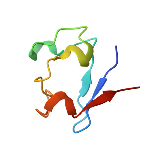
Understanding the structural origins of differences in reduction potentials is crucial to understanding how various electron transfer proteins modulate their reduction potentials and how they evolve for diverse functional roles. Here, the high-resolution structures of several Clostridium pasteurianum rubredoxin (Cp Rd) variants with changes in the vicinity of the redox site are reported in order to increase this understanding. Our crystal structures of [V44L] (at 1.8 A resolution), [V44A] (1.6 A), [V44G] (2.0 A) and [V44A, G45P] (1.5 A) Rd (all in their oxidized states) show that there is a gradual decrease in the distance between Fe and the amide nitrogen of residue 44 upon reduction in the size of the side chain of residue 44; the decrease occurs from leucine to valine, alanine or glycine and is accompanied by a gradual increase in their reduction potentials. Mutation of Cp Rd at position 44 also changes the hydrogen-bond distance between the amide nitrogen of residue 44 and the sulfur of cysteine 42 in a size-dependent manner. Our results suggest that residue 44 is an important determinant of Rd reduction potential in a manner dictated by side-chain size. Along with the electric dipole moment of the 43-44 peptide bond and the 44-42 NH--S type hydrogen bond, a modulation mechanism for solvent accessibility through residue 41 might regulate the redox reaction of the Rds.
Organizational Affiliation:
School of Molecular Biosciences, Washington State University, Pullman 99164-4660, USA.