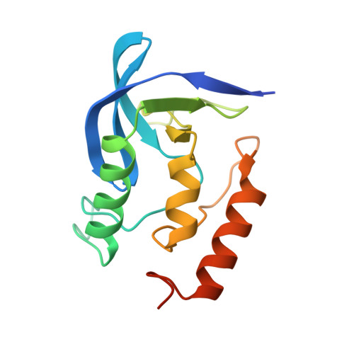
The crystal structure of staphylococcal nuclease refined at 1.7 A resolution.
Hynes, T.R., Fox, R.O.(1991) Proteins 10: 92-105
- PubMed: 1896431
- DOI: https://doi.org/10.1002/prot.340100203
- Primary Citation of Related Structures:
1STN - PubMed Abstract:
The crystal structure of staphylococcal nuclease has been determined to 1.7 A resolution with a final R-factor of 16.2% using stereochemically restrained Hendrickson-Konnert least-squares refinement. The structure reveals a number of conformational changes relative to the structure of the ternary complex of staphylococcal nuclease 1,2 bound with deoxythymidine-3',5'-diphosphate and Ca2+. Tyr-113 and Tyr-115, which pack against the nucleotide base in the nuclease complex, are rotated outward creating a more open binding pocket in the absence of nucleotide. The side chains of Ca2+ ligands Asp-21 and Asp-40 shift as does Glu-43, the proposed general base in the hydrolysis of the 5'-phosphodiester bond. The significance of some changes in the catalytic site is uncertain due to the intrusion of a symmetry related Lys-70 side chain which hydrogen bonds to both Asp-21 and Glu-43. The position of a flexible loop centered around residue 50 is altered, most likely due to conformational changes propagated from the Ca2+ site. The side chains of Arg-35, Lys-84, Tyr-85, and Arg-87, which hydrogen bond to the 3'- and 5'-phosphates of the nucleotide in the nuclease complex, are unchanged in conformation, with packing interactions with adjacent protein side chains sufficient to fix the geometry in the absence of ligand. The nuclease structure presented here, in combination with the stereochemically restrained refinement of the nuclease complex structure at 1.65 A, provides a wealth of structural information for the increasing number of studies using staphylococcal nuclease as a model system of protein structure and function.
Organizational Affiliation:
Department of Molecular Biophysics and Biochemistry, Yale University, New Haven, Connecticut 06511.