1SSF

Solution structure of the mouse 53BP1 fragment (residues 1463-1617)


Experimental Data Snapshot

  • Method: SOLUTION NMR
  • Conformers Calculated: 1000 
  • Conformers Submitted: 10 
  • Selection Criteria: back calculated data agree with experimental NOESY spectrum, structures with acceptable covalent geometry, structures with favorable non-bond energy, structures with the least restraint violations, structures with the lowest energy 

wwPDB Validation   3D Report Full Report


This is version 1.3 of the entry. See complete history


Literature

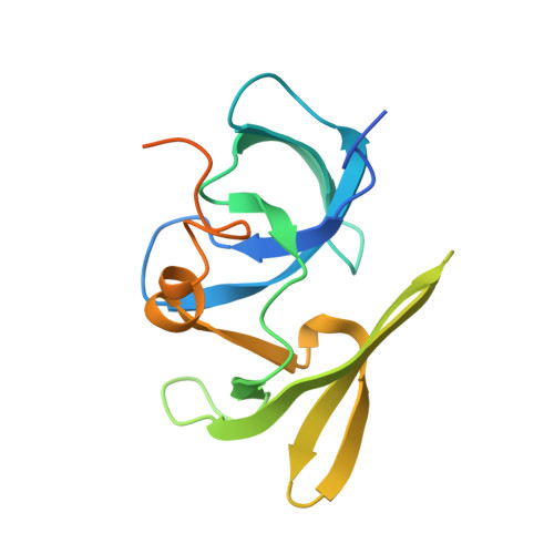
The Tudor Tandem of 53BP1; A New Structural Motif Involved in DNA and RG-Rich Peptide Binding

Charier, G.Couprie, J.Alpha-Bazin, B.Meyer, V.Quemeneur, E.Guerois, R.Callebaut, I.Gilquin, B.Zinn-Justin, S.

(2004) Structure 12: 1551-1562

  • DOI: https://doi.org/10.1016/j.str.2004.06.014
  • Primary Citation of Related Structures:  
    1SSF

  • PubMed Abstract: 

    53BP1 is a key transducer of the DNA damage checkpoint signal, which is required for phosphorylation of a subset of ATM substrates and p53 accumulation. After cell irradiation, the 53BP1 N-terminal region is phosphorylated. Its two C-terminal BRCT motifs interact with p53. Its central region is required and sufficient for 53BP1 foci formation at DNA strand breaks and for 53BP1 binding to the kinetochore. It contains an RG-rich segment and interacts with DNA in vitro. Here we show that the major globular domain of the 53BP1 central region adopts a new structural motif composed of two tightly packed Tudor domains and a C-terminal alpha helix. A unique surface essentially located on the first Tudor domain is involved in the binding to 53BP1 RG-rich sequence and to DNA, suggesting that the Tudor tandem can act as an adaptor mediating intramolecular as well as intermolecular protein-protein interactions and protein-nucleic acid associations.


  • Organizational Affiliation

    Département d'Ingénierie et d'Etudes des Protéines, 91191 Gif-sur-Yvette, France.


Macromolecules
Find similar proteins by:  (by identity cutoff)  |  3D Structure
Entity ID: 1
MoleculeChains Sequence LengthOrganismDetailsImage
Transformation related protein 53 binding protein 1156Mus musculusMutation(s): 0 
UniProt
Find proteins for P70399 (Mus musculus)
Explore P70399 
Go to UniProtKB:  P70399
Entity Groups  
Sequence Clusters30% Identity50% Identity70% Identity90% Identity95% Identity100% Identity
UniProt GroupP70399
Sequence Annotations
Expand
  • Reference Sequence
Experimental Data & Validation

Experimental Data

  • Method: SOLUTION NMR
  • Conformers Calculated: 1000 
  • Conformers Submitted: 10 
  • Selection Criteria: back calculated data agree with experimental NOESY spectrum, structures with acceptable covalent geometry, structures with favorable non-bond energy, structures with the least restraint violations, structures with the lowest energy 

Structure Validation

View Full Validation Report


Entry History 

Deposition Data

Revision History  (Full details and data files)

  • Version 1.0: 2004-09-14
    Type: Initial release
  • Version 1.1: 2008-04-29
    Changes: Version format compliance
  • Version 1.2: 2011-07-13
    Changes: Version format compliance
  • Version 1.3: 2022-03-02
    Changes: Data collection, Database references, Derived calculations