1SMN

IDENTIFICATION OF THE SERRATIA ENDONUCLEASE DIMER: STRUCTURAL BASIS AND IMPLICATIONS FOR CATALYSIS


Experimental Data Snapshot

  • Method: X-RAY DIFFRACTION
  • Resolution: 2.04 Å
  • R-Value Work: 0.168 
  • R-Value Observed: 0.168 

wwPDB Validation   3D Report Full Report


This is version 1.3 of the entry. See complete history


Literature

Identification of the Serratia endonuclease dimer: structural basis and implications for catalysis.

Miller, M.D.Krause, K.L.

(1996) Protein Sci 5: 24-33

  • DOI: https://doi.org/10.1002/pro.5560050104
  • Primary Citation of Related Structures:  
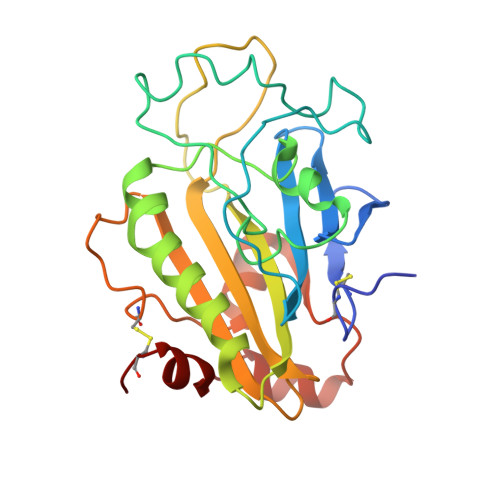
    1SMN

  • PubMed Abstract: 

    The Serratia endonuclease is an extracellularly secreted enzyme capable of cleaving both single- and double-stranded forms of DNA and RNA. It is the first member of a large class of related and usually dimeric endonucleases for which a structure is known. Using X-ray crystallography, the structure of monomer of this enzyme was reported by us previously (Miller MD et al., 1994, Nature Struct Biol 1:461-468). We now confirm the dimeric nature of this enzyme through light-scattering experiments and identify the physiologic dimer interface through crystal packing analysis. This dimerization occurs through an isologous twofold interaction localized to the carboxy-terminal subdomain of the enzyme. The dimer is a prolate ellipsoid with dimensions 30 A x 35 A x 90 A. The dimer interface is flat and contains four salt links, several hydrogen bonds, and nonpolar interactions. Buried water is prominent in this interface and it includes an unusual "cubic" water cluster. The position of the two active sites in the dimer suggests that they can act independently in their cleavage of DNA, but have a geometrical advantage in attacking substrate relative to the monomer.


  • Organizational Affiliation

    Department of Biochemical and Biophysical Sciences, University of Houston, Texas 77204-5934, USA.


Macromolecules
Find similar proteins by:  (by identity cutoff)  |  3D Structure
Entity ID: 1
MoleculeChains Sequence LengthOrganismDetailsImage
EXTRACELLULAR ENDONUCLEASE
A, B
245Serratia marcescensMutation(s): 0 
Gene Names: NUCA
EC: 3.1.30.2
UniProt
Find proteins for P13717 (Serratia marcescens)
Explore P13717 
Go to UniProtKB:  P13717
Entity Groups  
Sequence Clusters30% Identity50% Identity70% Identity90% Identity95% Identity100% Identity
UniProt GroupP13717
Sequence Annotations
Expand
  • Reference Sequence
Experimental Data & Validation

Experimental Data

  • Method: X-RAY DIFFRACTION
  • Resolution: 2.04 Å
  • R-Value Work: 0.168 
  • R-Value Observed: 0.168 
  • Space Group: P 21 21 2
Unit Cell:
Length ( Å )Angle ( ˚ )
a = 106.7α = 90
b = 74.5β = 90
c = 68.9γ = 90
Software Package:
Software NamePurpose
MADNESdata collection
PROCORdata collection
XSCALEdata scaling
X-PLORmodel building
X-PLORrefinement
MADNESdata reduction
PROCORdata reduction
X-PLORphasing

Structure Validation

View Full Validation Report


Entry History 

Deposition Data

Revision History  (Full details and data files)

  • Version 1.0: 1996-01-29
    Type: Initial release
  • Version 1.1: 2008-03-03
    Changes: Version format compliance
  • Version 1.2: 2011-07-13
    Changes: Version format compliance
  • Version 1.3: 2018-04-18
    Changes: Data collection