The refined crystal structure of subtilisin Carlsberg at 2.5 A resolution.
Neidhart, D.J., Petsko, G.A.(1988) Protein Eng 2: 271-276
- PubMed: 3150541
- DOI: https://doi.org/10.1093/protein/2.4.271
- Primary Citation of Related Structures:
1SBC - PubMed Abstract:
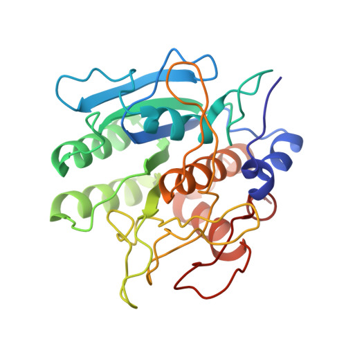
We report here the X-ray crystal structure of native subtilisin Carlsberg, solved at 2.5 A resolution by molecular replacement and refined by restrained least squares to a crystallographic residual (Formula see text): of 0.206. we compare this structure to the crystal structure of subtilisin BPN'. We find that, despite 82 amino acid substitutions and one deletion in subtilisin Carlsberg relative to subtilisin BPN', the structures of these enzymes are remarkably similar. We calculate an r.m.s. difference between equivalent alpha-carbon positions in subtilisin Carlsberg and subtilisin BPN' of only 0.55 A. This confirms previous reports of extensive structural homology between these two subtilisins based on X-ray crystal structures of the complex of eglin-c with subtilisin Carlsberg [McPhalen, C.A., Schnebli, H.P. and James, M.N.G. (1985) FEBS Lett., 188, 55; Bode, W., Papamokos, E. and Musil, D. (1987) Eur. J. Biochem., 166, 673-692]. In addition, we find that the native active sites of subtilisins Carlsberg and BPN' are virtually identical. While conservative substitutions at residues 217 and 156 may have subtle effects on the environments of substrate-binding sites S1' and S1 respectively, we find no obvious structural correlate for reports that subtilisins Carlsberg and BPN' differ in their recognition of model substrates. In particular, we find no evidence that the hydrophobic binding pocket S1 in subtilisin Carlsberg is 'deeper', 'narrower' or 'less polar' than the corresponding binding site in subtilisin BPN'.
Organizational Affiliation:
Department of Chemistry, Massachusetts Institute of Technology, Cambridge 02139.