1S4O

Crystal structure of yeast alpha1,2-mannosyltransferase Kre2p/Mnt1p: binary complex with GDP/Mn


Experimental Data Snapshot

  • Method: X-RAY DIFFRACTION
  • Resolution: 2.01 Å
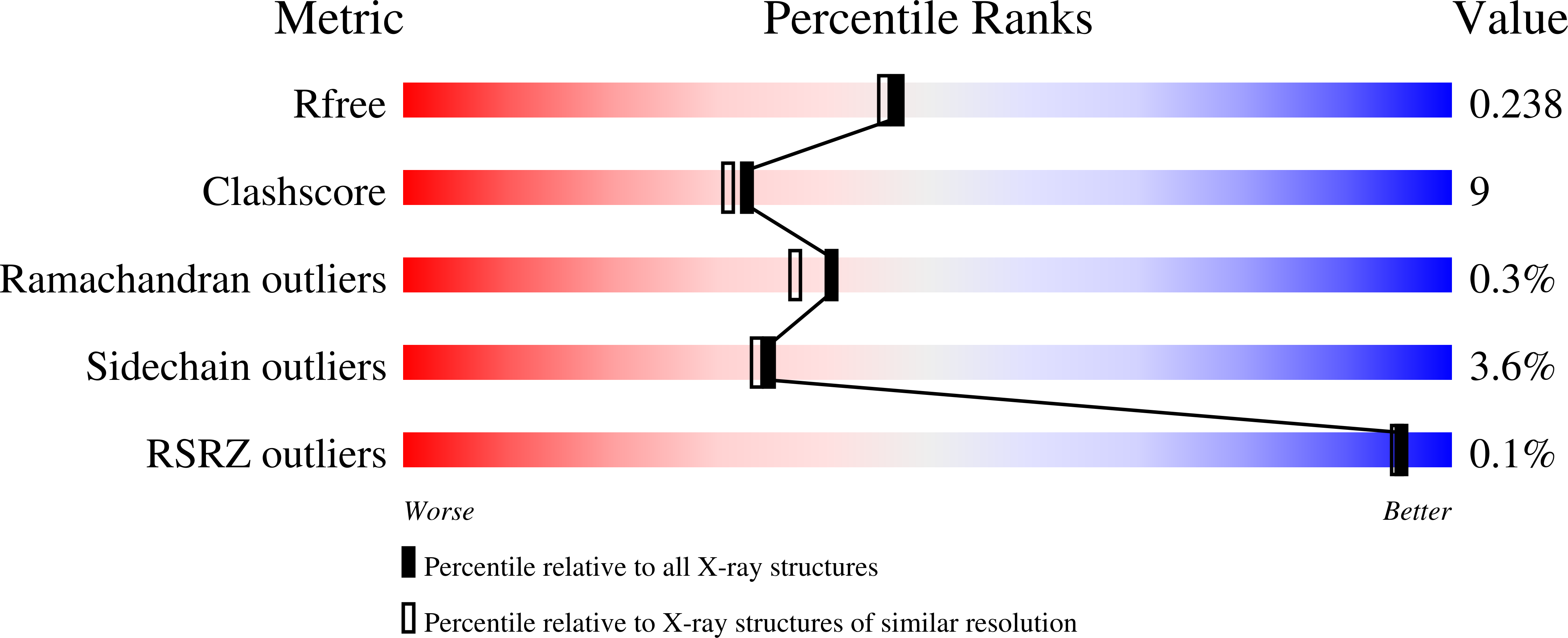
  • R-Value Free: 0.250 
  • R-Value Work: 0.197 
  • R-Value Observed: 0.197 

wwPDB Validation   3D Report Full Report


Ligand Structure Quality Assessment 


This is version 2.1 of the entry. See complete history


Literature

Structure of Kre2p/Mnt1p: A YEAST {alpha}1,2-MANNOSYLTRANSFERASE INVOLVED IN MANNOPROTEIN BIOSYNTHESIS

Lobsanov, Y.D.Romero, P.A.Sleno, B.Yu, B.Yip, P.Herscovics, A.Howell, P.L.

(2004) J Biol Chem 279: 17921-17931

  • DOI: https://doi.org/10.1074/jbc.M312720200
  • Primary Citation of Related Structures:  
    1S4N, 1S4O, 1S4P

  • PubMed Abstract: 

    Kre2p/Mnt1p is a Golgi alpha1,2-mannosyltransferase involved in the biosynthesis of Saccharomyces cerevisiae cell wall glycoproteins. The protein belongs to glycosyltransferase family 15, a member of which has been implicated in virulence of Candida albicans. We present the 2.0 A crystal structures of the catalytic domain of Kre2p/Mnt1p and its binary and ternary complexes with GDP/Mn(2+) and GDP/Mn(2+)/acceptor methyl-alpha-mannoside. The protein has a mixed alpha/beta fold similar to the glycosyltransferase-A (GT-A) fold. Although the GDP-mannose donor was used in the crystallization experiments and the GDP moiety is bound tightly to the active site, the mannose is not visible in the electron density. The manganese is coordinated by a modified DXD motif (EPD), with only the first glutamate involved in a direct interaction. The position of the donor mannose was modeled using the binary and ternary complexes of other GT-A enzymes. The C1" of the modeled donor mannose is within hydrogen-bonding distance of both the hydroxyl of Tyr(220) and the O2 of the acceptor mannose. The O2 of the acceptor mannose is also within hydrogen bond distance of the hydroxyl of Tyr(220). The structures, modeling, site-directed mutagenesis, and kinetic analysis suggest two possible catalytic mechanisms. Either a double-displacement mechanism with the hydroxyl of Tyr(220) as the potential nucleophile or alternatively, an S(N)i-like mechanism with Tyr(220) positioning the substrates for catalysis. The importance of Tyr(220) in both mechanisms is highlighted by a 3000-fold reduction in k(cat) in the Y220F mutant.


  • Organizational Affiliation

    Program in Structural Biology and Biochemistry, Research Institute, The Hospital for Sick Children, Toronto, Ontario M5G 1X8, Canada.


Macromolecules
Find similar proteins by:  (by identity cutoff)  |  3D Structure
Entity ID: 1
MoleculeChains Sequence LengthOrganismDetailsImage
Glycolipid 2-alpha-mannosyltransferase
A, B
348Saccharomyces cerevisiaeMutation(s): 0 
Gene Names: KRE2MNT1YDR483WD8035.26
EC: 2.4.1.131
UniProt
Find proteins for P27809 (Saccharomyces cerevisiae (strain ATCC 204508 / S288c))
Explore P27809 
Go to UniProtKB:  P27809
Entity Groups  
Sequence Clusters30% Identity50% Identity70% Identity90% Identity95% Identity100% Identity
UniProt GroupP27809
Sequence Annotations
Expand
  • Reference Sequence
Oligosaccharides

Help

Entity ID: 2
MoleculeChains Length2D Diagram Glycosylation3D Interactions
alpha-D-mannopyranose-(1-2)-alpha-D-mannopyranose-(1-3)-[alpha-D-mannopyranose-(1-3)-[alpha-D-mannopyranose-(1-6)]alpha-D-mannopyranose-(1-6)]beta-D-mannopyranose-(1-4)-2-acetamido-2-deoxy-alpha-D-glucopyranose-(1-4)-2-acetamido-2-deoxy-beta-D-glucopyranose
C
8N-Glycosylation
Glycosylation Resources
GlyTouCan:  G83314WY
GlyCosmos:  G83314WY
GlyGen:  G83314WY
Entity ID: 3
MoleculeChains Length2D Diagram Glycosylation3D Interactions
2-acetamido-2-deoxy-alpha-D-glucopyranose-(1-4)-2-acetamido-2-deoxy-beta-D-glucopyranose
D
2N-Glycosylation
Glycosylation Resources
GlyTouCan:  G07375KG
GlyCosmos:  G07375KG
GlyGen:  G07375KG
Small Molecules
Ligands 4 Unique
IDChains Name / Formula / InChI Key2D Diagram3D Interactions
GDP
Query on GDP

Download Ideal Coordinates CCD File 
G [auth A],
M [auth B]
GUANOSINE-5'-DIPHOSPHATE
C10 H15 N5 O11 P2
QGWNDRXFNXRZMB-UUOKFMHZSA-N
EPE
Query on EPE

Download Ideal Coordinates CCD File 
H [auth A],
N [auth B]
4-(2-HYDROXYETHYL)-1-PIPERAZINE ETHANESULFONIC ACID
C8 H18 N2 O4 S
JKMHFZQWWAIEOD-UHFFFAOYSA-N
MN
Query on MN

Download Ideal Coordinates CCD File 
E [auth A],
J [auth B]
MANGANESE (II) ION
Mn
WAEMQWOKJMHJLA-UHFFFAOYSA-N
CL
Query on CL

Download Ideal Coordinates CCD File 
F [auth A],
I [auth B],
K [auth B],
L [auth B]
CHLORIDE ION
Cl
VEXZGXHMUGYJMC-UHFFFAOYSA-M
Experimental Data & Validation

Experimental Data

  • Method: X-RAY DIFFRACTION
  • Resolution: 2.01 Å
  • R-Value Free: 0.250 
  • R-Value Work: 0.197 
  • R-Value Observed: 0.197 
  • Space Group: P 1 21 1
Unit Cell:
Length ( Å )Angle ( ˚ )
a = 60.694α = 90
b = 101.068β = 98.81
c = 62.036γ = 90
Software Package:
Software NamePurpose
CNSrefinement
CrystalCleardata reduction
d*TREKdata scaling
CNSphasing

Structure Validation

View Full Validation Report


Ligand Structure Quality Assessment 


Entry History 

Deposition Data

Revision History  (Full details and data files)

  • Version 1.0: 2004-05-04
    Type: Initial release
  • Version 1.1: 2008-04-29
    Changes: Version format compliance
  • Version 1.2: 2011-07-13
    Changes: Non-polymer description, Version format compliance
  • Version 2.0: 2020-07-29
    Type: Remediation
    Reason: Carbohydrate remediation
    Changes: Atomic model, Data collection, Database references, Derived calculations, Structure summary
  • Version 2.1: 2023-08-23
    Changes: Data collection, Database references, Refinement description, Structure summary