1S4I

Crystal structure of a SOD-like protein from Bacillus subtilis


Experimental Data Snapshot

  • Method: X-RAY DIFFRACTION
  • Resolution: 1.80 Å
  • R-Value Free: 0.258 
  • R-Value Work: 0.221 
  • R-Value Observed: 0.224 

wwPDB Validation   3D Report Full Report


This is version 1.2 of the entry. See complete history


Literature

A prokaryotic superoxide dismutase paralog lacking two Cu ligands: from largely unstructured in solution to ordered in the crystal.

Banci, L.Bertini, I.Calderone, V.Cramaro, F.Del Conte, R.Fantoni, A.Mangani, S.Quattrone, A.Viezzoli, M.S.

(2005) Proc Natl Acad Sci U S A 102: 7541-7546

  • DOI: https://doi.org/10.1073/pnas.0502450102
  • Primary Citation of Related Structures:  
    1S4I, 1U3N

  • PubMed Abstract: 

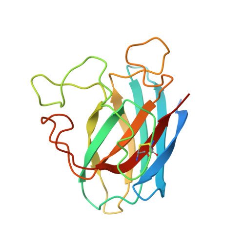
    Little is known about prokaryotic homologs of Cu,Zn superoxide dismutase (SOD), an enzyme highly conserved among eukaryotic species. In 138 Archaea and Bacteria genomes, 57 of these putative homologs were found, 11 of which lack at least one of the metal ligands. Both the solution and the crystal structures of the SOD-like protein from Bacillus subtilis, lacking two Cu ligands and found to be enzymatically inactive, were determined. In solution, the protein is monomeric. The available nuclear Overhauser effects, together with chemical-shift index values, allowed us to define and to recognize the typical Cu,Zn SOD Greek beta-barrel but with largely unstructured loops (which, therefore, sample a wide range of conformations). On the contrary, in the crystal structure (obtained in the presence of slight excess of Zn), the protein is well structured and organized in covalent dimers held by a symmetric bridge consisting of a Zn ion bound to an Asp-His dyad in a tetrahedral geometry. Couples of dimers held by hydrophobic interactions and H bonds are further organized in long chains. The order/disorder transition is discussed in terms of metal binding and physical state.


  • Organizational Affiliation

    Department of Chemistry, University of Florence, Via Luigi Sacconi 6, 50019 Sesto, Florence, Italy.


Macromolecules
Find similar proteins by:  (by identity cutoff)  |  3D Structure
Entity ID: 1
MoleculeChains Sequence LengthOrganismDetailsImage
superoxide dismutase-like protein yojMA [auth B],
B [auth A],
C,
D
175Bacillus subtilisMutation(s): 0 
Gene Names: YOJMBSU19400
EC: 1.15.1.1
UniProt
Find proteins for O31851 (Bacillus subtilis (strain 168))
Explore O31851 
Go to UniProtKB:  O31851
Entity Groups  
Sequence Clusters30% Identity50% Identity70% Identity90% Identity95% Identity100% Identity
UniProt GroupO31851
Sequence Annotations
Expand
  • Reference Sequence
Small Molecules
Ligands 2 Unique
IDChains Name / Formula / InChI Key2D Diagram3D Interactions
ZN
Query on ZN

Download Ideal Coordinates CCD File 
E [auth B]
G [auth B]
H [auth A]
J [auth A]
K [auth C]
E [auth B],
G [auth B],
H [auth A],
J [auth A],
K [auth C],
M [auth D]
ZINC ION
Zn
PTFCDOFLOPIGGS-UHFFFAOYSA-N
CL
Query on CL

Download Ideal Coordinates CCD File 
F [auth B],
I [auth A],
L [auth C],
N [auth D]
CHLORIDE ION
Cl
VEXZGXHMUGYJMC-UHFFFAOYSA-M
Experimental Data & Validation

Experimental Data

  • Method: X-RAY DIFFRACTION
  • Resolution: 1.80 Å
  • R-Value Free: 0.258 
  • R-Value Work: 0.221 
  • R-Value Observed: 0.224 
  • Space Group: P 1
Unit Cell:
Length ( Å )Angle ( ˚ )
a = 38.225α = 84.35
b = 61.108β = 76.02
c = 64.915γ = 90.42
Software Package:
Software NamePurpose
REFMACrefinement
MOSFLMdata reduction
CCP4data scaling
SHARPphasing

Structure Validation

View Full Validation Report


Entry History 

Deposition Data

Revision History  (Full details and data files)

  • Version 1.0: 2005-04-26
    Type: Initial release
  • Version 1.1: 2008-04-29
    Changes: Version format compliance
  • Version 1.2: 2011-07-13
    Changes: Derived calculations, Version format compliance