1S46

Covalent intermediate of the E328Q amylosucrase mutant


Experimental Data Snapshot

  • Method: X-RAY DIFFRACTION
  • Resolution: 2.20 Å
  • R-Value Free: 0.221 
  • R-Value Work: 0.184 
  • R-Value Observed: 0.184 

wwPDB Validation   3D Report Full Report


Ligand Structure Quality Assessment 


This is version 1.6 of the entry. See complete history


Literature

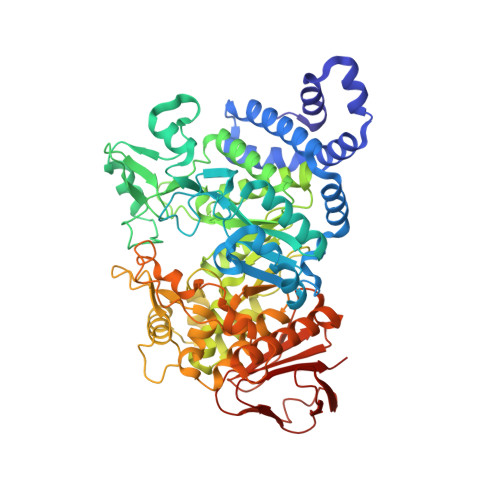
Crystal structure of the covalent intermediate of amylosucrase from Neisseria polysaccharea.

Jensen, M.H.Mirza, O.Albenne, C.Remaud-Simeon, M.Monsan, P.Gajhede, M.Skov, L.K.

(2004) Biochemistry 43: 3104-3110

  • DOI: https://doi.org/10.1021/bi0357762
  • Primary Citation of Related Structures:  
    1S46

  • PubMed Abstract: 

    The alpha-retaining amylosucrase from the glycoside hydrolase family 13 performs a transfer reaction of a glucosyl moiety from sucrose to an acceptor molecule. Amylosucrase has previously been shown to be able to use alpha-D-glucopyranosyl fluoride as a substrate, which suggested that it could also be used for trapping the reaction intermediate for crystallographic studies. In this paper, the crystal structure of the acid/base catalyst mutant, E328Q, with a covalently bound glucopyranosyl moiety is presented. Sucrose cocrystallized crystals were soaked with alpha-D-glucopyranosyl fluoride, which resulted in the trapping of a covalent intermediate in the active site of the enzyme. The structure is refined to a resolution of 2.2 A and showed that binding of the covalent intermediate resulted in a backbone movement of 1 A around the location of the nucleophile, Asp286. This structure reveals the first covalent intermediate of an alpha-retaining glycoside hydrolase where the glucosyl moiety is identical to the expected biologically relevant entity. Comparison to other enzymes with anticipated glucosylic covalent intermediates suggests that this structure is a representative model for such intermediates. Analysis of the active site shows how oligosaccharide binding disrupts the putative nucleophilic water binding site found in the hydrolases of the GH family 13. This reveals important parts of the structural background for the shift in function from hydrolase to transglycosidase seen in amylosucrase.


  • Organizational Affiliation

    Structural Biology Group, Department of Medicinal Chemistry, The Danish University of Pharmaceutical Sciences, Universitetsparken 2, DK-2100 Copenhagen, Denmark.


Macromolecules
Find similar proteins by:  (by identity cutoff)  |  3D Structure
Entity ID: 1
MoleculeChains Sequence LengthOrganismDetailsImage
amylosucrase628Neisseria polysacchareaMutation(s): 1 
EC: 2.4.1.4
UniProt
Find proteins for Q9ZEU2 (Neisseria polysaccharea)
Explore Q9ZEU2 
Go to UniProtKB:  Q9ZEU2
Entity Groups  
Sequence Clusters30% Identity50% Identity70% Identity90% Identity95% Identity100% Identity
UniProt GroupQ9ZEU2
Sequence Annotations
Expand
  • Reference Sequence
Small Molecules
Ligands 1 Unique
IDChains Name / Formula / InChI Key2D Diagram3D Interactions
BGC
Query on BGC

Download Ideal Coordinates CCD File 
B [auth A]beta-D-glucopyranose
C6 H12 O6
WQZGKKKJIJFFOK-VFUOTHLCSA-N
Experimental Data & Validation

Experimental Data

  • Method: X-RAY DIFFRACTION
  • Resolution: 2.20 Å
  • R-Value Free: 0.221 
  • R-Value Work: 0.184 
  • R-Value Observed: 0.184 
  • Space Group: P 21 21 2
Unit Cell:
Length ( Å )Angle ( ˚ )
a = 96.253α = 90
b = 116.63β = 90
c = 60.539γ = 90
Software Package:
Software NamePurpose
CNSrefinement
MAR345data collection
SCALEPACKdata scaling
CNSphasing

Structure Validation

View Full Validation Report


Ligand Structure Quality Assessment 


Entry History 

Deposition Data

Revision History  (Full details and data files)

  • Version 1.0: 2004-03-23
    Type: Initial release
  • Version 1.1: 2008-04-29
    Changes: Version format compliance
  • Version 1.2: 2011-07-13
    Changes: Version format compliance
  • Version 1.3: 2017-10-11
    Changes: Data collection, Refinement description
  • Version 1.4: 2018-01-24
    Changes: Database references
  • Version 1.5: 2020-07-29
    Type: Remediation
    Reason: Carbohydrate remediation
    Changes: Data collection, Database references, Derived calculations, Structure summary
  • Version 1.6: 2021-10-27
    Changes: Database references, Structure summary