1RRA

RIBONUCLEASE A FROM RATTUS NORVEGICUS (COMMON RAT)


Experimental Data Snapshot

  • Method: X-RAY DIFFRACTION
  • Resolution: 2.50 Å
  • R-Value Free: 0.277 
  • R-Value Work: 0.186 

wwPDB Validation   3D Report Full Report


This is version 1.3 of the entry. See complete history


Literature

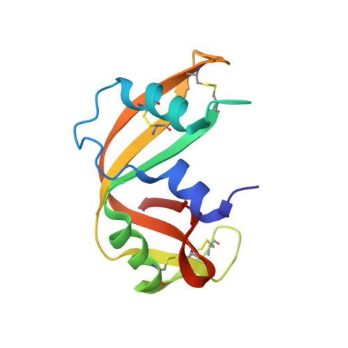
The crystal structure of recombinant rat pancreatic RNase A.

Gupta, V.Muyldermans, S.Wyns, L.Salunke, D.M.

(1999) Proteins 35: 1-12

  • DOI: https://doi.org/10.1002/(sici)1097-0134(19990401)35:1<1::aid-prot1>3.0.co;2-2
  • Primary Citation of Related Structures:  
    1RRA

  • PubMed Abstract: 

    The three-dimensional structure of rat pancreatic RNase A expressed in Escherichia coli was determined. The backbone conformations of certain critical loops are significantly different in this enzyme compared to its bovine counterpart. However, the core structure of rat RNase A is similar to that of the other members of the pancreatic ribonuclease family. The structural variations within a loop bordering the active site can be correlated with the subtle differences in the enzymatic activities of bovine and rat ribonucleases for different substrates. The most significant difference in the backbone conformation was observed in the loop 15-25. This loop incorporates the subtilisin cleavage site which is responsible for RNase A to RNase S conversion in the bovine enzyme. The rat enzyme does not get cleaved under identical conditions. Molecular docking of this region of the rat enzyme in the active site of subtilisin shows steric incompatibility, although the bovine pancreatic ribonuclease A appropriately fits into this active site. It is therefore inferred that the local conformation of the substrate governs the specificity of subtilisin.


  • Organizational Affiliation

    Structural Biology Unit, National Institute of Immunology, New Delhi, India.


Macromolecules
Find similar proteins by:  (by identity cutoff)  |  3D Structure
Entity ID: 1
MoleculeChains Sequence LengthOrganismDetailsImage
PROTEIN (RIBONUCLEASE)124Rattus norvegicusMutation(s): 0 
EC: 3.1.27.5
UniProt
Find proteins for P00684 (Rattus norvegicus)
Explore P00684 
Go to UniProtKB:  P00684
Entity Groups  
Sequence Clusters30% Identity50% Identity70% Identity90% Identity95% Identity100% Identity
UniProt GroupP00684
Sequence Annotations
Expand
  • Reference Sequence
Small Molecules
Ligands 1 Unique
IDChains Name / Formula / InChI Key2D Diagram3D Interactions
PO4
Query on PO4

Download Ideal Coordinates CCD File 
B [auth A]PHOSPHATE ION
O4 P
NBIIXXVUZAFLBC-UHFFFAOYSA-K
Experimental Data & Validation

Experimental Data

  • Method: X-RAY DIFFRACTION
  • Resolution: 2.50 Å
  • R-Value Free: 0.277 
  • R-Value Work: 0.186 
  • Space Group: P 31 2 1
Unit Cell:
Length ( Å )Angle ( ˚ )
a = 69.13α = 90
b = 69.13β = 90
c = 60.93γ = 120
Software Package:
Software NamePurpose
DENZOdata reduction
SCALEPACKdata scaling
AMoREphasing
X-PLORrefinement

Structure Validation

View Full Validation Report


Entry History 

Deposition Data

Revision History  (Full details and data files)

  • Version 1.0: 1998-12-09
    Type: Initial release
  • Version 1.1: 2008-04-26
    Changes: Version format compliance
  • Version 1.2: 2011-07-13
    Changes: Version format compliance
  • Version 1.3: 2023-08-23
    Changes: Data collection, Database references, Derived calculations, Refinement description