1RLM

Crystal Structure of ybiV from Escherichia coli K12


Experimental Data Snapshot

  • Method: X-RAY DIFFRACTION
  • Resolution: 1.90 Å
  • R-Value Free: 0.248 
  • R-Value Work: 0.219 
  • R-Value Observed: 0.219 

wwPDB Validation   3D Report Full Report


This is version 1.5 of the entry. See complete history


Literature

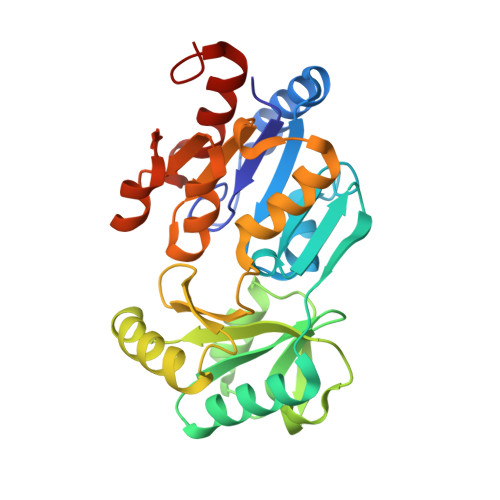
Ybiv from Escherichia coli K12 is a HAD phosphatase.

Roberts, A.Lee, S.Y.McCullagh, E.Silversmith, R.E.Wemmer, D.E.

(2005) Proteins 58: 790-801

  • DOI: https://doi.org/10.1002/prot.20267
  • Primary Citation of Related Structures:  
    1RLM, 1RLO, 1RLT

  • PubMed Abstract: 

    The protein YbiV from Escherichia coli K12 MG1655 is a hypothetical protein with sequence homology to the haloacid dehalogenase (HAD) superfamily of proteins. Although numerous members of this family have been identified, the functions of few are known. Using the crystal structure, sequence analysis, and biochemical assays, we have characterized YbiV as a HAD phosphatase. The crystal structure of YbiV reveals a two-domain protein, one with the characteristic HAD hydrolase fold, the other an inserted alpha/beta fold. In an effort to understand the mechanism, we also solved and report the structures of YbiV in complex with beryllofluoride (BeF3-) and aluminum trifluoride (AlF3), which have been shown to mimic the phosphorylated intermediate and transition state for hydrolysis, respectively, in analogy to other HAD phosphatases. Analysis of the structures reveals the substrate-binding cavity, which is hydrophilic in nature. Both structure and sequence homology indicate YbiV may be a sugar phosphatase, which is supported by biochemical assays that measured the release of free phosphate on a number of sugar-like substrates. We also investigated available genomic and functional data in an effort to determine the physiological substrate.


  • Organizational Affiliation

    Department of Chemistry, University of California, Berkeley, California 94720, USA.


Macromolecules
Find similar proteins by:  (by identity cutoff)  |  3D Structure
Entity ID: 1
MoleculeChains Sequence LengthOrganismDetailsImage
Phosphatase
A, B, C, D
271Escherichia coliMutation(s): 2 
Gene Names: YBIVB0822
EC: 3.1.3
UniProt
Find proteins for P75792 (Escherichia coli (strain K12))
Explore P75792 
Go to UniProtKB:  P75792
Entity Groups  
Sequence Clusters30% Identity50% Identity70% Identity90% Identity95% Identity100% Identity
UniProt GroupP75792
Sequence Annotations
Expand
  • Reference Sequence
Small Molecules
Ligands 2 Unique
IDChains Name / Formula / InChI Key2D Diagram3D Interactions
GOL
Query on GOL

Download Ideal Coordinates CCD File 
F [auth A]
G [auth A]
I [auth B]
K [auth C]
L [auth C]
F [auth A],
G [auth A],
I [auth B],
K [auth C],
L [auth C],
N [auth D]
GLYCEROL
C3 H8 O3
PEDCQBHIVMGVHV-UHFFFAOYSA-N
MG
Query on MG

Download Ideal Coordinates CCD File 
E [auth A],
H [auth B],
J [auth C],
M [auth D]
MAGNESIUM ION
Mg
JLVVSXFLKOJNIY-UHFFFAOYSA-N
Experimental Data & Validation

Experimental Data

  • Method: X-RAY DIFFRACTION
  • Resolution: 1.90 Å
  • R-Value Free: 0.248 
  • R-Value Work: 0.219 
  • R-Value Observed: 0.219 
  • Space Group: P 21 21 21
Unit Cell:
Length ( Å )Angle ( ˚ )
a = 71.823α = 90
b = 91.605β = 90
c = 186.563γ = 90
Software Package:
Software NamePurpose
CNSrefinement
DENZOdata reduction
SCALEPACKdata scaling
SOLVEphasing

Structure Validation

View Full Validation Report


Entry History 

Deposition Data

Revision History  (Full details and data files)

  • Version 1.0: 2004-12-07
    Type: Initial release
  • Version 1.1: 2008-04-29
    Changes: Version format compliance
  • Version 1.2: 2011-07-13
    Changes: Non-polymer description, Version format compliance
  • Version 1.3: 2014-11-12
    Changes: Structure summary
  • Version 1.4: 2021-10-27
    Changes: Database references, Derived calculations
  • Version 1.5: 2024-02-14
    Changes: Data collection