1RIO

Structure of bacteriophage lambda cI-NTD in complex with sigma-region4 of Thermus aquaticus bound to DNA


Experimental Data Snapshot

  • Method: X-RAY DIFFRACTION
  • Resolution: 2.30 Å
  • R-Value Free: 0.254 
  • R-Value Work: 0.216 
  • R-Value Observed: 0.218 

wwPDB Validation   3D Report Full Report


This is version 1.2 of the entry. See complete history


Literature

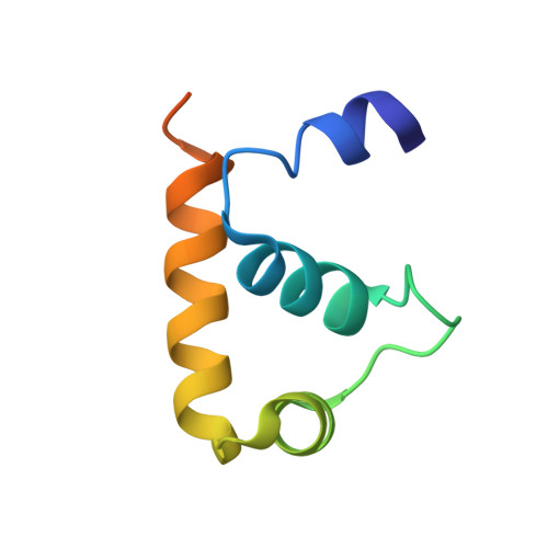
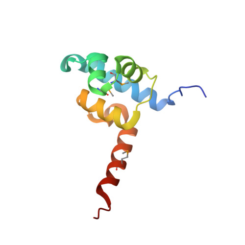
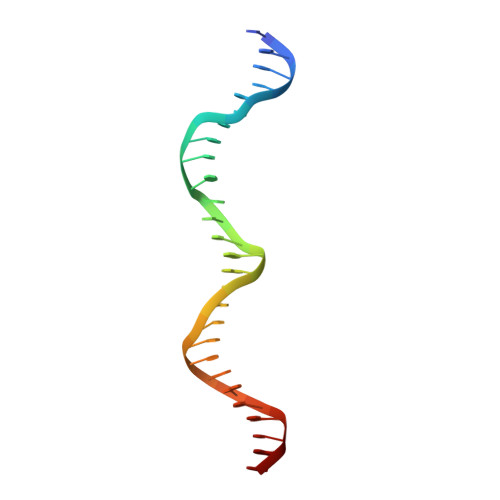
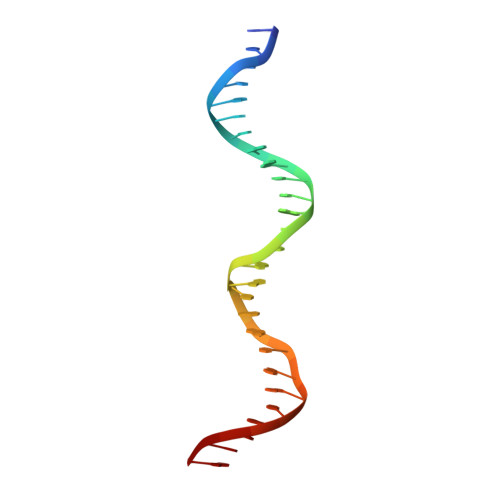
Structure of a ternary transcription activation complex.

Jain, D.Nickels, B.E.Sun, L.Hochschild, A.Darst, S.A.

(2004) Mol Cell 13: 45-53

  • DOI: https://doi.org/10.1016/s1097-2765(03)00483-0
  • Primary Citation of Related Structures:  
    1RIO

  • PubMed Abstract: 

    The cI protein of bacteriophage lambda (lambdacI) activates transcription by binding a DNA operator just upstream of the promoter and interacting with the RNA polymerase sigma subunit domain 4 (sigma(4)). We determined the crystal structure of the lambdacI/sigma(4)/DNA ternary complex at 2.3 A resolution. There are no conformational changes in either protein, which interact through an extremely small interface involving at most 6 amino acid residues. The interactions of the two proteins stabilize the binding of each protein to the DNA. The results provide insight into how activators can operate through a simple cooperative binding mechanism but affect different steps of the transcription initiation process.


  • Organizational Affiliation

    The Rockefeller University, 1230 York Avenue, New York, NY 10021, USA.


Macromolecules

Find similar proteins by:  (by identity cutoff)  |  3D Structure
Entity ID: 3
MoleculeChains Sequence LengthOrganismDetailsImage
sigma factor SigAC [auth H]73Thermus aquaticusMutation(s): 0 
UniProt
Find proteins for Q9EZJ8 (Thermus aquaticus)
Explore Q9EZJ8 
Go to UniProtKB:  Q9EZJ8
Entity Groups  
Sequence Clusters30% Identity50% Identity70% Identity90% Identity95% Identity100% Identity
UniProt GroupQ9EZJ8
Sequence Annotations
Expand
  • Reference Sequence
Find similar proteins by:  (by identity cutoff)  |  3D Structure
Entity ID: 4
MoleculeChains Sequence LengthOrganismDetailsImage
Repressor protein CID [auth A],
E [auth B]
98Lambdavirus lambdaMutation(s): 3 
Gene Names: CI
UniProt
Find proteins for P03034 (Escherichia phage lambda)
Explore P03034 
Go to UniProtKB:  P03034
Entity Groups  
Sequence Clusters30% Identity50% Identity70% Identity90% Identity95% Identity100% Identity
UniProt GroupP03034
Sequence Annotations
Expand
  • Reference Sequence
Find similar nucleic acids by:  (by identity cutoff)  |  3D Structure
Entity ID: 1
MoleculeChains LengthOrganismImage
27-MERA [auth U]27N/A
Sequence Annotations
Expand
  • Reference Sequence
Find similar nucleic acids by:  (by identity cutoff)  |  3D Structure
Entity ID: 2
MoleculeChains LengthOrganismImage
27-MERB [auth T]27N/A
Sequence Annotations
Expand
  • Reference Sequence
Small Molecules
Ligands 2 Unique
IDChains Name / Formula / InChI Key2D Diagram3D Interactions
MPD
Query on MPD

Download Ideal Coordinates CCD File 
G [auth A](4S)-2-METHYL-2,4-PENTANEDIOL
C6 H14 O2
SVTBMSDMJJWYQN-YFKPBYRVSA-N
CA
Query on CA

Download Ideal Coordinates CCD File 
F [auth A]CALCIUM ION
Ca
BHPQYMZQTOCNFJ-UHFFFAOYSA-N
Modified Residues  1 Unique
IDChains TypeFormula2D DiagramParent
MSE
Query on MSE
D [auth A],
E [auth B]
L-PEPTIDE LINKINGC5 H11 N O2 SeMET
Experimental Data & Validation

Experimental Data

  • Method: X-RAY DIFFRACTION
  • Resolution: 2.30 Å
  • R-Value Free: 0.254 
  • R-Value Work: 0.216 
  • R-Value Observed: 0.218 
  • Space Group: P 1 21 1
Unit Cell:
Length ( Å )Angle ( ˚ )
a = 47.256α = 90
b = 71.269β = 91.34
c = 77.199γ = 90
Software Package:
Software NamePurpose
REFMACrefinement
DENZOdata reduction
SCALEPACKdata scaling
MLPHAREphasing

Structure Validation

View Full Validation Report


Entry History 

Deposition Data

Revision History  (Full details and data files)

  • Version 1.0: 2004-01-27
    Type: Initial release
  • Version 1.1: 2008-04-29
    Changes: Version format compliance
  • Version 1.2: 2011-07-13
    Changes: Advisory, Version format compliance