1RHS

SULFUR-SUBSTITUTED RHODANESE


Experimental Data Snapshot

  • Method: X-RAY DIFFRACTION
  • Resolution: 1.36 Å
  • R-Value Free: 0.229 
  • R-Value Observed: 0.159 

wwPDB Validation   3D Report Full Report


This is version 1.2 of the entry. See complete history


Literature

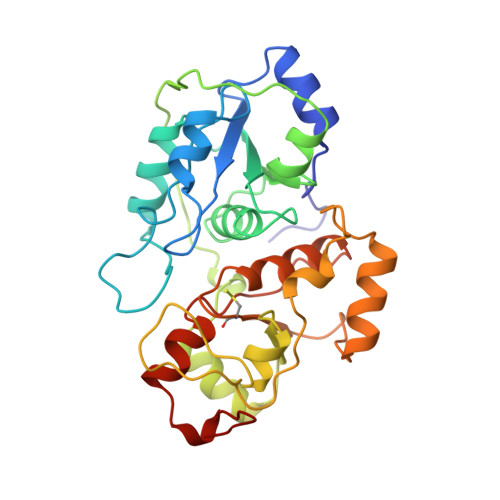
Structure of sulfur-substituted rhodanese at 1.36 A resolution.

Gliubich, F.Berni, R.Colapietro, M.Barba, L.Zanotti, G.

(1998) Acta Crystallogr D Biol Crystallogr 54: 481-486

  • DOI: https://doi.org/10.1107/s090744499701216x
  • Primary Citation of Related Structures:  
    1RHS

  • PubMed Abstract: 

    1.36 A resolution X-ray diffraction data have been recorded at 100 K for bovine liver sulfur-substituted rhodanese, using synchrotron radiation. The crystal structure has been refined anisotropically to a final R factor of 0.159 (Rfree = 0.229) for 53034 unique reflections. The model contains 2327 protein atoms and 407 solvent molecules, with a good geometry. The high resolution allows full details for helices, beta-sheets, tight turns and of all inter- and intramolecular interactions stabilizing the enzyme molecule to be given. The situation at the active site is described, particularly in regard to the network of hydrogen bonds made by Sgamma and Sdelta of the sulfur-substituted catalytic Cys247 and surrounding groups and solvent molecules. The replacement of the precipitant ammonium sulfate with cryoprotectants in the crystal-suspending medium led to the removal of the sulfate ion from the enzyme active site. Only limited changes of the enzyme structure have been found as a result of the drastic change in the crystal medium.


  • Organizational Affiliation

    Department of Organic Chemistry, University of Padova and Biopolymer Research Center, CNR, 35131, Padova, Italy.


Macromolecules
Find similar proteins by:  (by identity cutoff)  |  3D Structure
Entity ID: 1
MoleculeChains Sequence LengthOrganismDetailsImage
SULFUR-SUBSTITUTED RHODANESE296Bos taurusMutation(s): 0 
EC: 2.8.1.1
UniProt
Find proteins for P00586 (Bos taurus)
Explore P00586 
Go to UniProtKB:  P00586
Entity Groups  
Sequence Clusters30% Identity50% Identity70% Identity90% Identity95% Identity100% Identity
UniProt GroupP00586
Sequence Annotations
Expand
  • Reference Sequence
Small Molecules
Modified Residues  1 Unique
IDChains TypeFormula2D DiagramParent
CSS
Query on CSS
A
L-PEPTIDE LINKINGC3 H7 N O2 S2CYS
Experimental Data & Validation

Experimental Data

  • Method: X-RAY DIFFRACTION
  • Resolution: 1.36 Å
  • R-Value Free: 0.229 
  • R-Value Observed: 0.159 
  • Space Group: C 1 2 1
Unit Cell:
Length ( Å )Angle ( ˚ )
a = 154.364α = 90
b = 49.312β = 99.81
c = 41.676γ = 90
Software Package:
Software NamePurpose
SHELXL-93model building
SHELXL-93refinement
XDSdata reduction
MARSCALEdata scaling
SHELXL-93phasing

Structure Validation

View Full Validation Report


Entry History 

Deposition Data

Revision History  (Full details and data files)

  • Version 1.0: 1998-01-21
    Type: Initial release
  • Version 1.1: 2008-03-04
    Changes: Version format compliance
  • Version 1.2: 2011-07-13
    Changes: Version format compliance