Solution Structure of the C-terminal Antiparallel Coiled-coil Domain from Escherichia coli Osmosensor ProP.
Zoetewey, D.L., Tripet, B.P., Kutateladze, T.G., Overduin, M.J., Wood, J.M., Hodges, R.S.(2003) J Mol Biol 334: 1063-1076
- PubMed: 14643666
- DOI: https://doi.org/10.1016/j.jmb.2003.10.020
- Primary Citation of Related Structures:
1R48 - PubMed Abstract:
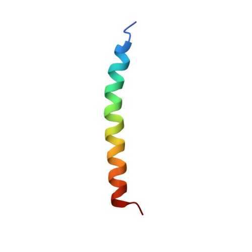
Bacteria respond to increasing medium osmolality by accumulating organic solutes that are compatible with cellular functions. Transporter ProP of Escherichia coli, a proton symporter and a member of the major facilitator superfamily, senses osmotic shifts and responds by importing osmolytes such as glycine betaine. ProP contains a cytoplasmic, C-terminal extension that is essential for its activity. A peptide corresponding to the C-terminal extension of ProP forms a homodimeric alpha-helical coiled-coil even though some of its heptad a positions are not occupied by hydrophobic amino acid residues. Unexpectedly, amino acid replacement R488I, occurring at a heptad a position, destabilized the coiled-coil formed by the ProP peptide and attenuated the response of the intact transporter to osmotic upshifts in vivo. Thus, ProP was proposed to dimerize via an antiparallel coiled-coil. We used nuclear magnetic resonance (NMR) spectroscopy to determine the structure of the synthetic peptide corresponding to residues 468-497 of ProP. This region did form an antiparallel coil-coil in which critical residue R488 specifies the antiparallel coiled-coil orientation by forming stabilizing salt-bridges. Charged residues (both acidic and basic) are clustered on the c/g surface of the coiled-coil whereas polar residues are distributed on the b/e surface. This causes the structure to be bent, in contrast to other known antiparallel coiled-coils (those from the hepatitis delta antigen (PDB ID code 1A92) and the bovine F(1) ATPase inhibitor protein (PDB ID code 1HF9)). The coiled-coil and its possible importance for osmosensing are discussed.
Organizational Affiliation:
Department of Biochemistry and Molecular Genetics, University of Colorado Health Sciences Center, Denver, CO 80262, USA.