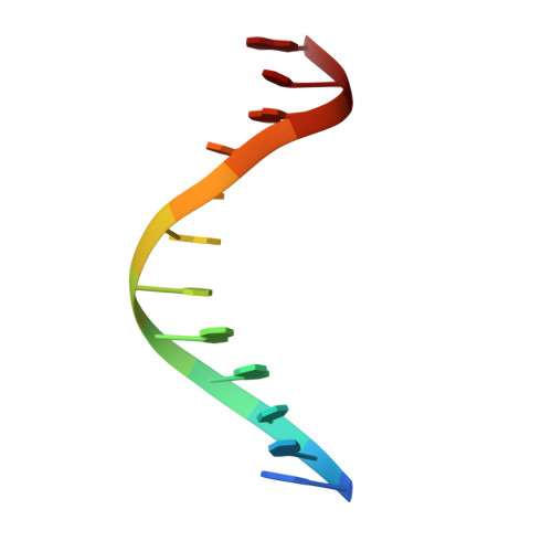
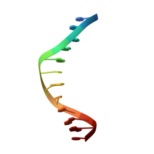
Base-pairing shift in the major groove of (CA)n tracts by B-DNA crystal structures.
Timsit, Y., Vilbois, E., Moras, D.(1991) Nature 354: 167-170
- PubMed: 1944598
- DOI: https://doi.org/10.1038/354167a0
- Primary Citation of Related Structures:
1QP5, 330D - PubMed Abstract:
the crystal packing of the B-DNA dodecamer d(ACCG-GCGCCACA).d(TGTGGCGCCGGT) is characterized by the reciprocal fit of double helices with specific base-backbone interactions in the major groove. Cooling the crystals below -10 degrees C stabilizes a new conformational state with a long-range sequence-dependent one-step shift in the major-groove base pairing. The tilt of the bases leads to the disruption of the Watson-Crick pairing in the major groove and to the formation of interactions with the 5' neighbour of their complement. This alteration propagates along the helical axis over more than half a turn. As a result, the molecular structure is normal when seen from the minor groove side and mismatched in the major groove. Comparison with a parent isomorphous dodecamer structure corresponding to the codon 10-13 of the c-Ha-ras proto-oncogene show that this new structural feature is sequence dependent and clearly favoured by (CA)n tracts. As(CA)n tracts of DNA are involved both in recombination and in transcription, this new recognition pattern should be considered in the analysis of the various processes involving the reading of the genetic information.
Organizational Affiliation:
Laboratoire de Cristallographie Biologique, Institut de Biologie Moléculaire et Cellulaire, Strasbourg, France.


