Stability of a Structural Scaffold Upon Activity Transfer : X-Ray Structure of a Three Fingers Chimeric Protein.
Le Du, M.H., Ricciardi, A., Khayati, M., Menez, R., Boulain, J.C., Menez, A., Ducancel, F.(2000) J Mol Biol 296: 1017
- PubMed: 10686100
- DOI: https://doi.org/10.1006/jmbi.2000.3510
- Primary Citation of Related Structures:
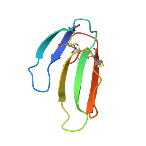
1QM7 - PubMed Abstract:
Fasciculin 2 and toxin alpha proteins belong to the same structural family of three-fingered snake toxins. They act on different targets, but in each case the binding region involves residues from loops I and II. The superimposition of the two structures suggests that these functional regions correspond to structurally distinct zones. Loop I, half of loop II and the C-terminal residue of fasciculin 2 were therefore transferred into the toxin alpha. The inhibition constant of the resulting chimera is only 15-fold lower than that of fasciculin 2, and as expected the potency of binding to the toxin alpha target has been lost. In order to understand the structure-function relationship between the chimera and its "parent" molecules, we solved its structure by X-ray crystallography. The protein crystallized in space group P3(1)21 with a=b=58.5 A, and c=62.3 A. The crystal structure was solved by molecular replacement and refined to 2.1 A resolution. The structure belongs to the three-fingered snake toxin family with a core of four disulphide bridges from which emerge the three loops I, II and III. Superimposition of the chimera on fasciculin 2 or toxin alpha revealed an overall fold intermediate between those of the two parent molecules. The regions corresponding to toxin alpha and to fasciculin 2 retained their respective geometries. In addition, the chimera protein displayed a structural behaviour similar to that of fasciculin 2, i.e. dimerization in the crystal structure of fasciculin 2, and the geometry of the region that binds to acetylcholinesterase. In conclusion, this structure shows that the chimera retains the general structural characteristics of three-fingered toxins, and the structural specificity of the transferred function.
Organizational Affiliation:
Département d'Ingénierie et d'Etude des Protéines, CE Saclay, Gif-sur-Yvette Cedex, 91191, France. mhledu@cea.fr