1QB2

CRYSTAL STRUCTURE OF THE CONSERVED SUBDOMAIN OF HUMAN PROTEIN SRP54M AT 2.1A RESOLUTION: EVIDENCE FOR THE MECHANISM OF SIGNAL PEPTIDE BINDING


Experimental Data Snapshot

  • Method: X-RAY DIFFRACTION
  • Resolution: 2.10 Å
  • R-Value Free: 0.318 
  • R-Value Work: 0.245 

wwPDB Validation   3D Report Full Report


This is version 1.4 of the entry. See complete history


Literature

Crystal structure of the conserved subdomain of human protein SRP54M at 2.1 A resolution: evidence for the mechanism of signal peptide binding.

Clemons Jr., W.M.Gowda, K.Black, S.D.Zwieb, C.Ramakrishnan, V.

(1999) J Mol Biol 292: 697-705

  • DOI: https://doi.org/10.1006/jmbi.1999.3090
  • Primary Citation of Related Structures:  
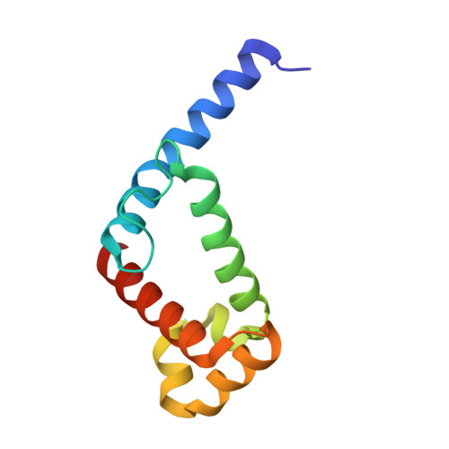
    1QB2

  • PubMed Abstract: 

    Protein SRP54 is an integral part of the mammalian signal recognition particle (SRP), a cytosolic ribonucleoprotein complex which associates with ribosomes and serves to recognize, bind, and transport proteins destined for the membrane or secretion. The methionine-rich M-domain of protein SRP54 (SRP54M) binds the SRP RNA and the signal peptide as the nascent protein emerges from the ribosome. A focal point of this critical cellular function is the detailed understanding of how different hydrophobic signal peptides are recognized efficiently and transported specifically, despite considerable variation in sequence. We have solved the crystal structure of a conserved functional subdomain of the human SRP54 protein (hSRP54m) at 2.1 A resolution showing a predominantly alpha helical protein with a large fraction of the structure available for binding. RNA binding is predicted to occur in the vicinity of helices 4 to 6. The N-terminal helix extends significantly from the core of the structure into a large but constricted hydrophobic groove of an adjacent molecule, thus revealing molecular details of possible interactions between alpha helical signal peptides and human SRP54.


  • Organizational Affiliation

    Department of Biochemistry, The University of Utah, School of Medicine, 50 North Medical Drive, Salt Lake City, UT 84132, USA.


Macromolecules
Find similar proteins by:  (by identity cutoff)  |  3D Structure
Entity ID: 1
MoleculeChains Sequence LengthOrganismDetailsImage
HUMAN SIGNAL RECOGNITION PARTICLE 54 KD PROTEIN
A, B
109Homo sapiensMutation(s): 0 
UniProt & NIH Common Fund Data Resources
Find proteins for P61011 (Homo sapiens)
Explore P61011 
Go to UniProtKB:  P61011
PHAROS:  P61011
GTEx:  ENSG00000100883 
Entity Groups  
Sequence Clusters30% Identity50% Identity70% Identity90% Identity95% Identity100% Identity
UniProt GroupP61011
Sequence Annotations
Expand
  • Reference Sequence
Experimental Data & Validation

Experimental Data

  • Method: X-RAY DIFFRACTION
  • Resolution: 2.10 Å
  • R-Value Free: 0.318 
  • R-Value Work: 0.245 
  • Space Group: P 21 21 21
Unit Cell:
Length ( Å )Angle ( ˚ )
a = 28.91α = 90
b = 61.34β = 90
c = 129.22γ = 90
Software Package:
Software NamePurpose
SOLVEphasing
X-PLORrefinement
MADNESSdata reduction
SCALEPACKdata scaling

Structure Validation

View Full Validation Report


Entry History 

Deposition Data

Revision History  (Full details and data files)

  • Version 1.0: 1999-10-20
    Type: Initial release
  • Version 1.1: 2008-04-27
    Changes: Version format compliance
  • Version 1.2: 2011-07-13
    Changes: Version format compliance
  • Version 1.3: 2017-10-04
    Changes: Refinement description
  • Version 1.4: 2024-02-14
    Changes: Data collection, Database references