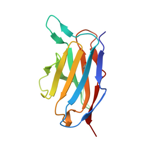
1QAC

CHANGE IN DIMERIZATION MODE BY REMOVAL OF A SINGLE UNSATISFIED POLAR RESIDUE


Experimental Data Snapshot

  • Method: X-RAY DIFFRACTION
  • Resolution: 1.80 Å
  • R-Value Free: 0.226 
  • R-Value Work: 0.193 

wwPDB Validation   3D Report Full Report


This is version 1.4 of the entry. See complete history


Literature

Change in dimerization mode by removal of a single unsatisfied polar residue located at the interface.

Pokkuluri, P.R.Cai, X.Johnson, G.Stevens, F.J.Schiffer, M.

(2000) Protein Sci 9: 1852-1855

  • DOI: https://doi.org/10.1110/ps.9.9.1852
  • Primary Citation of Related Structures:  
    1QAC, 5LVE

  • PubMed Abstract: 

    The importance of unsatisfied hydrogen bonding potential on protein-protein interaction was studied. Two alternate modes of dimerization (conventional and flipped form) of an immunoglobulin light chain variable domain (V(L)) were previously identified. In the flipped form, interface residue Gln89 would have an unsatisfied hydrogen bonding potential. Removal of this Gln should render the flipped dimer as the more favorable quaternary form. High resolution crystallographic studies of the Q89A and Q89L mutants show, as we predicted, that these proteins indeed form flipped dimers with very similar interfaces. A small cavity is present in the Q89A mutant that is reflected in the approximately 100 times lower association constant than found for the Q89L mutant. The association constant of Q89A and Q89L proteins (4 x 10(6) M(-1) and >10(8) M(-1)) are 10- and 1,000-fold higher than that of the wild-type protein that forms conventional dimers clearly showing the energetic reasons for the flipped dimer formation.


  • Organizational Affiliation

    Argonne National Laboratory, Biosciences Division, Illinois 60439, USA.


Macromolecules
Find similar proteins by:  (by identity cutoff)  |  3D Structure
Entity ID: 1
MoleculeChains Sequence LengthOrganismDetailsImage
IMMUNOGLOBULIN LIGHT CHAIN VARIABLE DOMAIN
A, B
114Homo sapiensMutation(s): 0 
UniProt & NIH Common Fund Data Resources
Find proteins for P06312 (Homo sapiens)
Explore P06312 
Go to UniProtKB:  P06312
PHAROS:  P06312
GTEx:  ENSG00000211598 
Entity Groups  
Sequence Clusters30% Identity50% Identity70% Identity90% Identity95% Identity100% Identity
UniProt GroupP06312
Sequence Annotations
Expand
  • Reference Sequence
Experimental Data & Validation

Experimental Data

  • Method: X-RAY DIFFRACTION
  • Resolution: 1.80 Å
  • R-Value Free: 0.226 
  • R-Value Work: 0.193 
  • Space Group: P 1 21 1
Unit Cell:
Length ( Å )Angle ( ˚ )
a = 35.3α = 90
b = 83.6β = 109
c = 42.8γ = 90
Software Package:
Software NamePurpose
SCALEPACKdata scaling
AMoREphasing
CNSrefinement

Structure Validation

View Full Validation Report


Entry History 

Deposition Data

Revision History  (Full details and data files)

  • Version 1.0: 2000-02-23
    Type: Initial release
  • Version 1.1: 2008-04-27
    Changes: Version format compliance
  • Version 1.2: 2011-07-13
    Changes: Version format compliance
  • Version 1.3: 2017-10-04
    Changes: Refinement description
  • Version 1.4: 2023-08-16
    Changes: Data collection, Database references, Refinement description