1Q91

Crystal structure of human mitochondrial deoxyribonucleotidase in complex with the inhibitor DPB-T


Experimental Data Snapshot

  • Method: X-RAY DIFFRACTION
  • Resolution: 1.60 Å
  • R-Value Free: 0.206 
  • R-Value Work: 0.186 
  • R-Value Observed: 0.186 

wwPDB Validation   3D Report Full Report


Ligand Structure Quality Assessment 


This is version 1.3 of the entry. See complete history


Literature

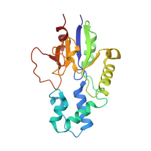
Crystal structures of the mitochondrial deoxyribonucleotidase in complex with two specific inhibitors

Rinaldo-Matthis, A.Rampazzo, C.Balzarini, J.Reichard, P.Bianchi, V.Nordlund, P.

(2004) Mol Pharmacol 65: 860-867

  • DOI: https://doi.org/10.1124/mol.65.4.860
  • Primary Citation of Related Structures:  
    1Q91, 1Q92

  • PubMed Abstract: 

    Monophosphate nucleotidases are enzymes that dephosphorylate nucleotides to their corresponding nucleosides. They play potentially important roles in controlling the activation of nucleotide-based drugs targeted against viral infections or cancer cells. The human mitochondrial deoxyribonucleotidase (dNT-2) dephosphorylates thymidine and deoxyuridine monophosphates. We describe the high resolution structures of the dNT-2 enzyme in complex with two potent nucleoside phosphonate inhibitors, (S)-1-[2'-deoxy-3',5'-O-(1-phosphono) benzylidene-beta-d-threo-pentofuranosyl]thymine (DPB-T) at 1.6-A resolution and (+/-)-1-trans-(2-phosphonomethoxycyclopentyl)uracil (PMcP-U) at 1.4-A resolution. The mixed competitive inhibitor DPB-T and the competitive inhibitor PMcP-U both bind in the active site of dNT-2 but in distinctly different binding modes, explaining their different kinetics of inhibition. The pyrimidine part of the inhibitors binds with very similar hydrogen bond interactions to the protein but with their phosphonate moieties in different binding sites compared with each other and to the previously determined position of phosphate bound to dNT-2. Together, these phosphate/phosphonate binding sites describe what might constitute a functionally relevant phosphate entrance tunnel to the active site. The structures of the inhibitors in complex with dNT-2, being the first such complexes of any nucleotidase, might provide important information for the design of more specific inhibitors to control the activation of nucleotide-based drugs.


  • Organizational Affiliation

    Department of Biochemistry and Biophysics, Stockholm University, Stockholm, Sweden.


Macromolecules
Find similar proteins by:  (by identity cutoff)  |  3D Structure
Entity ID: 1
MoleculeChains Sequence LengthOrganismDetailsImage
5(3)-deoxyribonucleotidase197Homo sapiensMutation(s): 0 
EC: 3.1.3.5
UniProt & NIH Common Fund Data Resources
Find proteins for Q9NPB1 (Homo sapiens)
Explore Q9NPB1 
Go to UniProtKB:  Q9NPB1
PHAROS:  Q9NPB1
GTEx:  ENSG00000205309 
Entity Groups  
Sequence Clusters30% Identity50% Identity70% Identity90% Identity95% Identity100% Identity
UniProt GroupQ9NPB1
Sequence Annotations
Expand
  • Reference Sequence
Small Molecules
Ligands 3 Unique
IDChains Name / Formula / InChI Key2D Diagram3D Interactions
DPB
Query on DPB

Download Ideal Coordinates CCD File 
D [auth A](S)-1-[2'-DEOXY-3',5'-O-(1-PHOSPHONO)BENZYLIDENE-B-D-THREO-PENTOFURANOSYL]THYMINE
C17 H19 N2 O8 P
WTZFKHNHHRPQOU-WSMBLCCSSA-N
PO4
Query on PO4

Download Ideal Coordinates CCD File 
B [auth A]PHOSPHATE ION
O4 P
NBIIXXVUZAFLBC-UHFFFAOYSA-K
MG
Query on MG

Download Ideal Coordinates CCD File 
C [auth A]MAGNESIUM ION
Mg
JLVVSXFLKOJNIY-UHFFFAOYSA-N
Binding Affinity Annotations 
IDSourceBinding Affinity
DPB PDBBind:  1Q91 Ki: 7.00e+4 (nM) from 1 assay(s)
Binding MOAD:  1Q91 Ki: 7.00e+4 (nM) from 1 assay(s)
Experimental Data & Validation

Experimental Data

  • Method: X-RAY DIFFRACTION
  • Resolution: 1.60 Å
  • R-Value Free: 0.206 
  • R-Value Work: 0.186 
  • R-Value Observed: 0.186 
  • Space Group: P 43 21 2
Unit Cell:
Length ( Å )Angle ( ˚ )
a = 73.651α = 90
b = 73.651β = 90
c = 106.83γ = 90
Software Package:
Software NamePurpose
CNSrefinement
DENZOdata reduction
SCALEPACKdata scaling
CNSphasing

Structure Validation

View Full Validation Report


Ligand Structure Quality Assessment 


Entry History 

Deposition Data

Revision History  (Full details and data files)

  • Version 1.0: 2004-04-20
    Type: Initial release
  • Version 1.1: 2008-04-29
    Changes: Version format compliance
  • Version 1.2: 2011-07-13
    Changes: Version format compliance
  • Version 1.3: 2023-10-25
    Changes: Data collection, Database references, Derived calculations, Refinement description