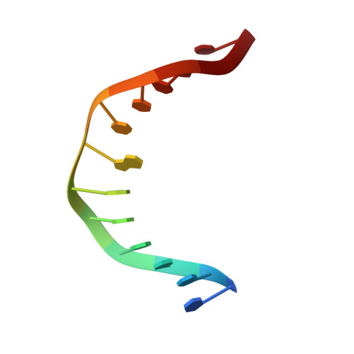
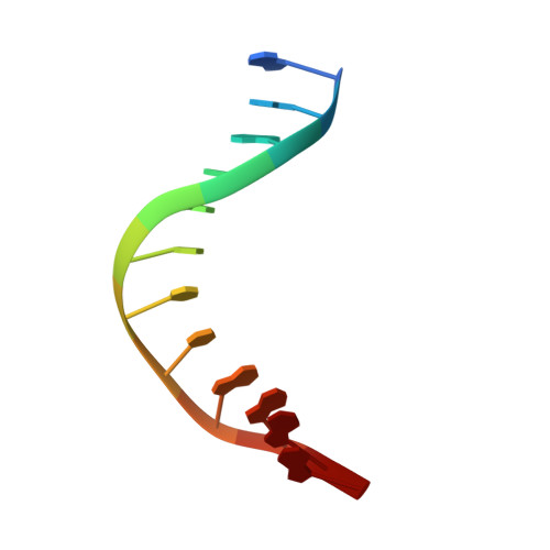
NMR solution structure of an oxaliplatin 1,2-d(GG) intrastrand cross-link in a DNA dodecamer duplex
Wu, Y., Pradhan, P., Havener, J., Boysen, G., Swenberg, J.A., Campbel, S.L., Chaney, S.G.(2004) J Mol Biol 341: 1251-1269
- PubMed: 15321720
- DOI: https://doi.org/10.1016/j.jmb.2004.06.066
- Primary Citation of Related Structures:
1PG9, 1PGC - PubMed Abstract:
We have determined, at high resolution, the NMR solution structure of an oxaliplatin-GG DNA dodecamer in the AGGC sequence context by 2D NMR studies. Homonuclear assignment strategies resulted in unambiguous assignment of 203 out of 249 protons, which corresponds to assignment of approximately 81% of the protons. Assignments of H5' and H5" protons were tentative due to resonance overlap. The structure of the oxaliplatin duplex was calculated using the program CNS with a simulated annealing protocol. A total of 510 experimental restraints were employed in the structure calculation. Of 20 calculated structures, the 15 with the lowest energy were accepted as a family. The RMSD of the 15 lowest energy structures was 0.68 A, indicating good structural convergence. The theoretical NOESY spectrum obtained by back-calculation from the final average structure showed excellent agreement with the experimental data, indicating that the final structure was in good agreement with the experimental NMR data. Significant conformational differences were observed between the oxaliplatin-GG 12-mer DNA we studied and all previous solution structures of cisplatin-GG DNA duplexes. For example, the oxaliplatin-GG adduct shows much less distortion at the AG base-pair step than the cisplatin-GG adducts. In addition, the oxaliplatin-GG structure also has a narrow minor groove and an overall axis bend of about 31 degrees, both of which are very different from the recent NMR structures for the cisplatin-GG adducts. These structural differences may explain some of the biological differences between oxaliplatin- and cisplatin-GG adducts.
Organizational Affiliation:
Department of Biochemistry and Biophysics, School of Medicine, University of North Carolina, Chapel Hill, NC 27599-7260, USA.