Cobalt(II), nickel(II) and zinc(II) do not bind to intra-helical N(7) guanine positions in the B-form crystal structure of d(GGCGCC)
Labiuk, S.L., Delbaere, L.T., Lee, J.S.(2003) J Biol Inorg Chem 8: 715-720
- PubMed: 14505075
- DOI: https://doi.org/10.1007/s00775-003-0473-4
- Primary Citation of Related Structures:
1P24, 1P25, 1P26 - PubMed Abstract:
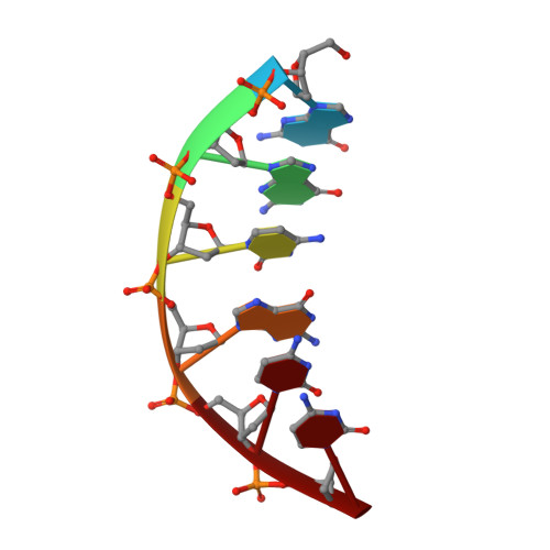
Three novel X-ray crystal structures for the DNA hexamer d(GGCGCC) in the B-form complexed to divalent cobalt, nickel and zinc ions have been determined to a resolution of 2.9-3.0 A. The structures were isomorphous and had five DNA strands and five metal cations per asymmetric unit. In all three cases, divalent metal cations were coordinated only to the terminal guanine residue at the N(7) position, with no metal ions binding to non-terminal guanine positions. Water molecules bound to the metal cations interacted with neighboring guanine residues 3' to the ones to which the cations were coordinated, affecting the propeller twist. Even though DNA occupied only about 35% of the unit cell volume, it is interesting that the few interactions involving the metal cations were sufficient to stabilize the crystal lattice. As well as lending support to the proposal that these metals do not coordinate to B-DNA in a stable manner, the results presented here also extend the crystallographic evidence for this phenomenon to the GGC and CGC sequences for all three metal cations.
Organizational Affiliation:
Department of Biochemistry, University of Saskatchewan, 107 Wiggins Road, S7N 5E5, Saskatoon, SK, Canada.