Monoclinic guanidinoacetate methyltransferase and gadolinium ion-binding characteristics.
Komoto, J., Takata, Y., Yamada, T., Konishi, K., Ogawa, H., Gomi, T., Fujioka, M., Takusagawa, F.(2003) Acta Crystallogr D Biol Crystallogr 59: 1589-1596
- PubMed: 12925789
- DOI: https://doi.org/10.1107/s0907444903014719
- Primary Citation of Related Structures:
1P1B, 1P1C - PubMed Abstract:
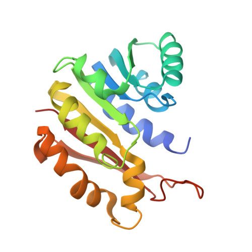
Guanidinoacetate methyltransferase (GAMT) is the enzyme that catalyzes the last step of creatine biosynthesis. The enzyme is found in abundance in the livers of all vertebrates. Recombinant rat liver GAMT truncated at amino acid 37 from the N-terminus has been crystallized with S-adenosylhomocysteine (SAH) in a monoclinic modification and the crystal structure has been determined at 2.8 A resolution. There are two dimers in the crystallographic asymmetric unit. Each dimer has non-crystallographic twofold symmetry and is related to the other dimer by pseudo-4(3) symmetry along the crystallographic b axis. The overall structure of GAMT crystallized in the monoclinic modification is quite similar to the structure observed in the tetragonal modification [Komoto et al. (2002), J. Mol. Biol. 320, 223-235], with the exception of the loop containing Tyr136. In the monoclinic modification, the loops in three of the four subunits have a catalytically unfavorable conformation and the loop of the fourth subunit has a catalytically favorable conformation as observed in the crystals of the tetragonal modification. From the structures in the monoclinic and tetragonal modifications, we can explain why the Y136F mutant enzyme retains considerable catalytic activity while the Y136V mutant enzyme loses the catalytic activity. The crystal structure of a Gd derivative of the tetragonal modification has also been determined. By comparing the Gd-derivative structure with the native structures in the tetragonal and the monoclinic modifications, useful characteristic features of Gd-ion binding for application in protein crystallography have been observed. Gd ions can bind to proteins without changing the native protein structures and Gd atoms produce strong anomalous dispersion signals from Cu Kalpha radiation; however, Gd-ion binding to protein requires a relatively specific geometry.
Organizational Affiliation:
Department of Molecular Biosciences, University of Kansas, 1200 Sunnyside Avenue, Lawrence, KS 66045-7534, USA.