1OZ6

X-ray structure of acidic phospholipase A2 from Indian saw-scaled viper (Echis carinatus) with a potent platelet aggregation inhibitory activity


Experimental Data Snapshot

  • Method: X-RAY DIFFRACTION
  • Resolution: 2.60 Å
  • R-Value Free: 0.257 
  • R-Value Work: 0.192 
  • R-Value Observed: 0.207 

wwPDB Validation   3D Report Full Report


This is version 1.3 of the entry. See complete history


Literature

Structure of an acidic phospholipase A2 from Indian saw-scaled viper (Echis carinatus) at 2.6 A resolution reveals a novel intermolecular interaction.

Jasti, J.Paramasivam, M.Srinivasan, A.Singh, T.P.

(2004) Acta Crystallogr D Biol Crystallogr 60: 66-72

  • DOI: https://doi.org/10.1107/s090744490302208x
  • Primary Citation of Related Structures:  
    1OZ6

  • PubMed Abstract: 

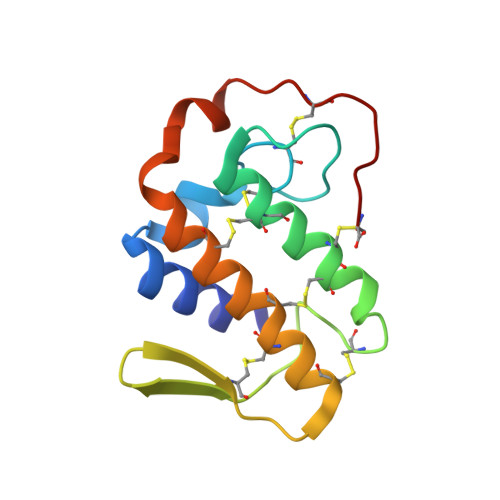
    The crystal structure of an acidic phospholipase A(2) from the venom of Echis carinatus (saw-scaled viper; scPLA(2)) has been determined at 2.6 A resolution and refined to a crystallographic R factor of 0.192. Although the overall structure of scPLA(2) is essentially similar to those of other group II acidic PLA(2)s from different species, it shows unique features in several parts. Particularly noteworthy is the C-terminal part, which folds differently to those of other group II PLA(2)s. This part is considered to be responsible for inhibition of the platelet-aggregation activity. The calcium-binding loop is tightly organized with sevenfold coordination. Another striking feature of scPLA(2) is the involvement of Asn79 O(delta1) of a symmetry-related molecule in a coordination linkage with Ca(2+) of the calcium-binding loop. This is the first observation of an internal metal ion participating in an intermolecular interaction. The beta-wing of a molecule is deeply inserted into the hydrophobic channel of another molecule and forms several intermolecular interactions. This results in the formation of an infinite chain of molecules. These chains are stacked in an antiparallel arrangement in the crystals.


  • Organizational Affiliation

    Department of Biophysics, All India Institute of Medical Sciences, New Delhi 110 029, India.


Macromolecules
Find similar proteins by:  (by identity cutoff)  |  3D Structure
Entity ID: 1
MoleculeChains Sequence LengthOrganismDetailsImage
phospholipase A2120Echis carinatusMutation(s): 0 
EC: 3.1.1.4
Membrane Entity: Yes 
UniProt
Find proteins for Q7T3S7 (Echis carinatus)
Explore Q7T3S7 
Go to UniProtKB:  Q7T3S7
Entity Groups  
Sequence Clusters30% Identity50% Identity70% Identity90% Identity95% Identity100% Identity
UniProt GroupQ7T3S7
Sequence Annotations
Expand
  • Reference Sequence
Small Molecules
Ligands 1 Unique
IDChains Name / Formula / InChI Key2D Diagram3D Interactions
CA
Query on CA

Download Ideal Coordinates CCD File 
B [auth A]CALCIUM ION
Ca
BHPQYMZQTOCNFJ-UHFFFAOYSA-N
Experimental Data & Validation

Experimental Data

  • Method: X-RAY DIFFRACTION
  • Resolution: 2.60 Å
  • R-Value Free: 0.257 
  • R-Value Work: 0.192 
  • R-Value Observed: 0.207 
  • Space Group: P 21 21 2
Unit Cell:
Length ( Å )Angle ( ˚ )
a = 63.963α = 90
b = 57.876β = 90
c = 33.682γ = 90
Software Package:
Software NamePurpose
REFMACrefinement
DENZOdata reduction
SCALEPACKdata scaling
MOLREPphasing

Structure Validation

View Full Validation Report


Entry History 

Deposition Data

Revision History  (Full details and data files)

  • Version 1.0: 2003-12-30
    Type: Initial release
  • Version 1.1: 2008-04-29
    Changes: Version format compliance
  • Version 1.2: 2011-07-13
    Changes: Version format compliance
  • Version 1.3: 2023-10-25
    Changes: Data collection, Database references, Derived calculations, Refinement description