Determination of the solution structures of conantokin-G and conantokin-T by CD and NMR spectroscopy.
Skjaerbaek, N., Nielsen, K.J., Lewis, R.J., Alewood, P., Craik, D.J.(1997) J Biol Chem 272: 2291-2299
- PubMed: 8999936
- DOI: https://doi.org/10.1074/jbc.272.4.2291
- Primary Citation of Related Structures:
1ONT, 1ONU - PubMed Abstract:
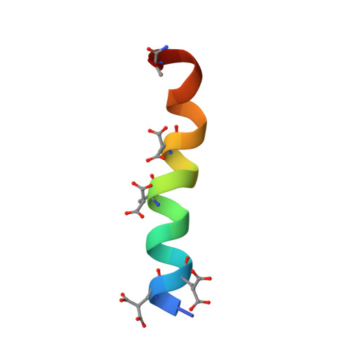
Conantokin-G and conantokin-T are two paralytic polypeptide toxins originally isolated from the venom of the fish-hunting cone snails of the genus Conus. Conantokin-G and conantokin-T are the only naturally occurring peptidic compounds which possess N-methyl-D-aspartate receptor antagonist activity, produced by a selective non-competitive antagonism of polyamine responses. They are also structurally unusual in that they contain a disproportionately large number of acid labile post-translational gamma-carboxyglutamic acid (Gla) residues. Although no precise structural information has previously been published for these peptides, early spectroscopic measurements have indicated that both conantokin-G and conantokin-T form alpha-helical structures, although there is some debate whether the presence of calcium ions is required for these peptides to adopt this fold. We now report a detailed structural study of synthetic conantokin-G and conantokin-T in a range of solution conditions using CD and 1H NMR spectroscopy. The three-dimensional structures of conantokin-T and conantokin-G were calculated from 1H NMR-derived distance and dihedral restraints. Both conantokins were found to contain a mixture of alpha- and 310 helix, that give rise to curved and straight helical conformers. Conantokin-G requires the presence of divalent cations (Zn2+, Ca2+, Cu2+, or Mg2+) to form a stable alpha-helix, while conantokin-T adopts a stable alpha-helical structure in aqueous conditions, in the presence or absence of divalent cations (Zn2+, Ca2+, Cu2+, or Mg2+).
Organizational Affiliation:
The Centre for Drug Design and Development, The University of Queensland, Brisbane, Qld 4072, Australia.