1OD7

N-terminal of Sialoadhesin in complex with Me-a-9-N-(naphthyl-2-carbonyl)-amino-9-deoxy-Neu5Ac (NAP compound)


Experimental Data Snapshot

  • Method: X-RAY DIFFRACTION
  • Resolution: 3.00 Å
  • R-Value Free: 0.272 
  • R-Value Work: 0.192 
  • R-Value Observed: 0.192 

wwPDB Validation   3D Report Full Report


Ligand Structure Quality Assessment 


This is version 1.3 of the entry. See complete history


Literature

Structure-Guided Design of Sialic Acid-Based Siglec Inhibitors and Crystallographic Analysis in Complex with Sialoadhesin

Zaccai, N.R.Maenaka, K.Maenaka, T.Crocker, P.R.Brossmer, R.Kelm, S.Jones, E.Y.

(2003) Structure 11: 557

  • DOI: https://doi.org/10.1016/s0969-2126(03)00073-x
  • Primary Citation of Related Structures:  
    1OD7, 1OD9, 1ODA

  • PubMed Abstract: 

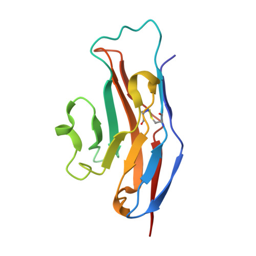
    The Siglec family of receptors mediates cell surface interactions through recognition of sialylated glycoconjugates. The crystal structure of the N-terminal immunoglobulin-like domain of the Siglec sialoadhesin (SnD1) in complex with 2,3-sialyllactose has informed the design of sialic acid analogs (sialosides) that bind Siglecs with significantly enhanced affinities and specificities. Binding assays against sialoadhesin (Sn; Siglec-1), CD22 (Siglec-2), and MAG (Siglec-4) show a 10- to 300-fold reduction in IC(50) values (relative to methyl-alpha-Neu5Ac) for three sialosides bearing aromatic group modifications of the glycerol side chain: Me-alpha-9-N-benzoyl-amino-9-deoxy-Neu5Ac (BENZ), Me-alpha-9-N-(naphthyl-2-carbonyl)-amino-9-deoxy-Neu5Ac (NAP), and Me-alpha-9-N-(biphenyl-4-carbonyl)-amino-9-deoxy-Neu5Ac (BIP). Crystal structures of these sialosides in complex with SnD1 suggest explanations for the differences in specificity and affinity, providing further ideas for compound design of physiological and potentially therapeutic relevance.


  • Organizational Affiliation

    CR-UK Receptor Structure Research Group, Division of Structural Biology, The Henry Wellcome Building for Genomic Medicine, Roosevelt Drive, Headington, Oxford OX3 7BN, United Kingdom.


Macromolecules
Find similar proteins by:  (by identity cutoff)  |  3D Structure
Entity ID: 1
MoleculeChains Sequence LengthOrganismDetailsImage
SIALOADHESIN119Mus musculusMutation(s): 0 
UniProt & NIH Common Fund Data Resources
Find proteins for Q62230 (Mus musculus)
Explore Q62230 
Go to UniProtKB:  Q62230
IMPC:  MGI:99668
Entity Groups  
Sequence Clusters30% Identity50% Identity70% Identity90% Identity95% Identity100% Identity
UniProt GroupQ62230
Sequence Annotations
Expand
  • Reference Sequence
Small Molecules
Ligands 1 Unique
IDChains Name / Formula / InChI Key2D Diagram3D Interactions
SUW
Query on SUW

Download Ideal Coordinates CCD File 
B [auth A]ME-A-9-N-(NAPHTHYL-2-CARBONYL)-AMINO-9-DEOXY-NEU5AC
C23 H28 N2 O9
NCJJDBXUFQPTHE-GCIBVHORSA-N
Experimental Data & Validation

Experimental Data

  • Method: X-RAY DIFFRACTION
  • Resolution: 3.00 Å
  • R-Value Free: 0.272 
  • R-Value Work: 0.192 
  • R-Value Observed: 0.192 
  • Space Group: P 43 21 2
Unit Cell:
Length ( Å )Angle ( ˚ )
a = 64.866α = 90
b = 64.866β = 90
c = 64.051γ = 90
Software Package:
Software NamePurpose
CNSrefinement
DENZOdata reduction
SCALEPACKdata scaling
AMoREphasing

Structure Validation

View Full Validation Report


Ligand Structure Quality Assessment 


Entry History 

Deposition Data

Revision History  (Full details and data files)

  • Version 1.0: 2003-05-16
    Type: Initial release
  • Version 1.1: 2011-05-08
    Changes: Version format compliance
  • Version 1.2: 2011-07-13
    Changes: Version format compliance
  • Version 1.3: 2023-12-13
    Changes: Data collection, Database references, Derived calculations, Other, Refinement description