1O9I

Crystal structure of the Y42F mutant of manganese catalase from Lactobacillus plantarum at 1.33A resolution


Experimental Data Snapshot

  • Method: X-RAY DIFFRACTION
  • Resolution: 1.33 Å
  • R-Value Free: 0.145 
  • R-Value Work: 0.115 
  • R-Value Observed: 0.117 

wwPDB Validation   3D Report Full Report


This is version 1.6 of the entry. See complete history


Literature

Outer Sphere Mutagenesis of Lactobacillus Plantarum Manganese Catalase Disrupts the Cluster Core. Mechanistic Implications.

Whittaker, M.M.Barynin, V.V.Igarashi, T.Whittaker, J.W.

(2003) Eur J Biochem 270: 1102

  • DOI: https://doi.org/10.1046/j.1432-1033.2003.03459.x
  • Primary Citation of Related Structures:  
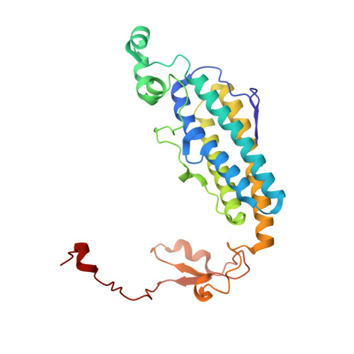
    1O9I

  • PubMed Abstract: 

    X-ray crystallography of the nonheme manganese catalase from Lactobacillus plantarum (LPC) [Barynin, V.V., Whittaker, M.M., Antonyuk, S.V., Lamzin, V.S., Harrison, P.M., Artymiuk, P.J. & Whittaker, J.W. (2001) Structure9, 725-738] has revealed the structure of the dimanganese redox cluster together with its protein environment. The oxidized [Mn(III)Mn(III)] cluster is bridged by two solvent molecules (oxo and hydroxo, respectively) together with a micro 1,3 bridging glutamate carboxylate and is embedded in a web of hydrogen bonds involving an outer sphere tyrosine residue (Tyr42). A novel homologous expression system has been developed for production of active recombinant LPC and Tyr42 has been replaced by phenylalanine using site-directed mutagenesis. Spectroscopic and structural studies indicate that disruption of the hydrogen-bonded web significantly perturbs the active site in Y42F LPC, breaking one of the solvent bridges and generating an 'open' form of the dimanganese cluster. Two of the metal ligands adopt alternate conformations in the crystal structure, both conformers having a broken solvent bridge in the dimanganese core. The oxidized Y42F LPC exhibits strong optical absorption characteristic of high spin Mn(III) in low symmetry and lower coordination number. MCD and EPR measurements provide complementary information defining a ferromagnetically coupled electronic ground state for a cluster containing a single solvent bridge, in contrast to the diamagnetic ground state found for the native cluster containing a pair of solvent bridges. Y42F LPC has less than 5% of the catalase activity and much higher Km for H2O2 ( approximately 1.4 m) at neutral pH than WT LPC, although the activity is slightly restored at high pH where the cluster is converted to a diamagnetic form. These studies provide new insight into the contribution of the outer sphere tyrosine to the stability of the dimanganese cluster and the role of the solvent bridges in catalysis by dimanganese catalases.


  • Organizational Affiliation

    Department of Environmental and Biomolecular Systems, OGI School of Science and Engineering at OHSU, Oregon, USA. jim@bmb.ogi.edu


Macromolecules
Find similar proteins by:  (by identity cutoff)  |  3D Structure
Entity ID: 1
MoleculeChains Sequence LengthOrganismDetailsImage
Manganese catalase
A, B, C, D, E
A, B, C, D, E, F
266Lactiplantibacillus plantarumMutation(s): 0 
EC: 1.11.1.6
UniProt
Find proteins for P60355 (Lactiplantibacillus plantarum)
Explore P60355 
Go to UniProtKB:  P60355
Entity Groups  
Sequence Clusters30% Identity50% Identity70% Identity90% Identity95% Identity100% Identity
UniProt GroupP60355
Sequence Annotations
Expand
  • Reference Sequence
Small Molecules
Ligands 5 Unique
IDChains Name / Formula / InChI Key2D Diagram3D Interactions
MES
Query on MES

Download Ideal Coordinates CCD File 
DA [auth E],
EA [auth E],
JA [auth F],
K [auth A],
KA [auth F]
2-(N-MORPHOLINO)-ETHANESULFONIC ACID
C6 H13 N O4 S
SXGZJKUKBWWHRA-UHFFFAOYSA-N
MN3
Query on MN3

Download Ideal Coordinates CCD File 
AA [auth E]
FA [auth F]
G [auth A]
GA [auth F]
H [auth A]
AA [auth E],
FA [auth F],
G [auth A],
GA [auth F],
H [auth A],
N [auth B],
O [auth B],
R [auth C],
S [auth C],
V [auth D],
W [auth D],
Z [auth E]
MANGANESE (III) ION
Mn
MMIPFLVOWGHZQD-UHFFFAOYSA-N
CA
Query on CA

Download Ideal Coordinates CCD File 
BA [auth E]
HA [auth F]
I [auth A]
P [auth B]
T [auth C]
BA [auth E],
HA [auth F],
I [auth A],
P [auth B],
T [auth C],
X [auth D]
CALCIUM ION
Ca
BHPQYMZQTOCNFJ-UHFFFAOYSA-N
NA
Query on NA

Download Ideal Coordinates CCD File 
L [auth A],
M [auth A]
SODIUM ION
Na
FKNQFGJONOIPTF-UHFFFAOYSA-N
O
Query on O

Download Ideal Coordinates CCD File 
CA [auth E]
IA [auth F]
J [auth A]
Q [auth B]
U [auth C]
CA [auth E],
IA [auth F],
J [auth A],
Q [auth B],
U [auth C],
Y [auth D]
OXYGEN ATOM
O
XLYOFNOQVPJJNP-UHFFFAOYSA-N
Experimental Data & Validation

Experimental Data

  • Method: X-RAY DIFFRACTION
  • Resolution: 1.33 Å
  • R-Value Free: 0.145 
  • R-Value Work: 0.115 
  • R-Value Observed: 0.117 
  • Space Group: P 1 21 1
Unit Cell:
Length ( Å )Angle ( ˚ )
a = 73.49α = 90
b = 95.27β = 106.55
c = 105γ = 90
Software Package:
Software NamePurpose
REFMACrefinement
DENZOdata reduction
SCALEPACKdata scaling

Structure Validation

View Full Validation Report


Entry History 

Deposition Data

Revision History  (Full details and data files)

  • Version 1.0: 2003-12-18
    Type: Initial release
  • Version 1.1: 2011-05-08
    Changes: Version format compliance
  • Version 1.2: 2011-07-13
    Changes: Version format compliance
  • Version 1.3: 2018-06-20
    Changes: Data collection, Database references, Source and taxonomy, Structure summary
  • Version 1.4: 2019-01-30
    Changes: Data collection, Experimental preparation
  • Version 1.5: 2019-05-22
    Changes: Data collection, Experimental preparation
  • Version 1.6: 2023-12-13
    Changes: Data collection, Database references, Derived calculations, Refinement description