1O8V

The crystal structure of Echinococcus granulosus fatty-acid-binding protein 1


Experimental Data Snapshot

  • Method: X-RAY DIFFRACTION
  • Resolution: 1.60 Å
  • R-Value Free: 0.214 
  • R-Value Work: 0.172 
  • R-Value Observed: 0.174 

wwPDB Validation   3D Report Full Report


Ligand Structure Quality Assessment 


This is version 1.4 of the entry. See complete history


Literature

The Crystal Structure of Echinococcus Granulosus Fatty-Acid-Binding Protein 1

Jakobsson, E.Alvite, G.Bergfors, T.Esteves, A.Kleywegt, G.J.

(2003) Biochim Biophys Acta 1649: 40

  • DOI: https://doi.org/10.1016/s1570-9639(03)00151-1
  • Primary Citation of Related Structures:  
    1O8V

  • PubMed Abstract: 

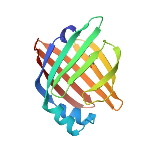
    We describe the 1.6 A crystal structure of the fatty-acid-binding protein EgFABP1 from the parasitic platyhelminth Echinococcus granulosus. E. granulosus causes hydatid disease, which is a major zoonosis. EgFABP1 has been implicated in the acquisition, storage, and transport of lipids, and may be important to the organism since it is incapable of synthesising most of its lipids de novo. Moreover, EgFABP1 is a promising candidate for a vaccine against hydatid disease. The crystal structure reveals that EgFABP1 has the expected 10-stranded beta-barrel fold typical of the family of intracellular lipid-binding proteins, and that it is structurally most similar to P2 myelin protein. We describe the comparison of the crystal structure of EgFABP1 with these proteins and with an older homology model for EgFABP1. The electron density reveals the presence of a bound ligand inside the cavity, which we have interpreted as palmitic acid. The carboxylate group of the fatty acid interacts with the protein's P2 motif, consisting of a conserved triad R em leader R-x-Y. The hydrophobic tail of the ligand assumes a fairly flat, U-shaped conformation and has relatively few interactions with the protein.We discuss some of the structural implications of the crystal structure of EgFABP1 for related platyhelminthic FABPs.


  • Organizational Affiliation

    Department of Cell and Molecular Biology, Uppsala University, Biomedical Centre, Box 596, SE-751 24, Uppsala, Sweden.


Macromolecules
Find similar proteins by:  (by identity cutoff)  |  3D Structure
Entity ID: 1
MoleculeChains Sequence LengthOrganismDetailsImage
FATTY ACID BINDING PROTEIN HOMOLOG134Echinococcus granulosusMutation(s): 0 
UniProt
Find proteins for Q02970 (Echinococcus granulosus)
Explore Q02970 
Go to UniProtKB:  Q02970
Entity Groups  
Sequence Clusters30% Identity50% Identity70% Identity90% Identity95% Identity100% Identity
UniProt GroupQ02970
Sequence Annotations
Expand
  • Reference Sequence
Small Molecules
Ligands 1 Unique
IDChains Name / Formula / InChI Key2D Diagram3D Interactions
PLM (Subject of Investigation/LOI)
Query on PLM

Download Ideal Coordinates CCD File 
B [auth A]PALMITIC ACID
C16 H32 O2
IPCSVZSSVZVIGE-UHFFFAOYSA-N
Modified Residues  1 Unique
IDChains TypeFormula2D DiagramParent
CSO
Query on CSO
A
L-PEPTIDE LINKINGC3 H7 N O3 SCYS
Experimental Data & Validation

Experimental Data

  • Method: X-RAY DIFFRACTION
  • Resolution: 1.60 Å
  • R-Value Free: 0.214 
  • R-Value Work: 0.172 
  • R-Value Observed: 0.174 
  • Space Group: P 1 21 1
Unit Cell:
Length ( Å )Angle ( ˚ )
a = 28.714α = 90
b = 54.834β = 100.34
c = 38.691γ = 90
Software Package:
Software NamePurpose
REFMACrefinement
DENZOdata reduction
SCALEPACKdata scaling
AMoREphasing

Structure Validation

View Full Validation Report


Ligand Structure Quality Assessment 


Entry History 

Deposition Data

Revision History  (Full details and data files)

  • Version 1.0: 2003-06-19
    Type: Initial release
  • Version 1.1: 2011-05-08
    Changes: Version format compliance
  • Version 1.2: 2011-07-13
    Changes: Version format compliance
  • Version 1.3: 2019-10-09
    Changes: Author supporting evidence, Data collection, Derived calculations, Other, Structure summary
  • Version 1.4: 2023-12-13
    Changes: Data collection, Database references, Refinement description