1O8B

Structure of Escherichia coli ribose-5-phosphate isomerase, RpiA, complexed with arabinose-5-phosphate.


Experimental Data Snapshot

  • Method: X-RAY DIFFRACTION
  • Resolution: 1.25 Å
  • R-Value Free: 0.240 
  • R-Value Work: 0.224 
  • R-Value Observed: 0.224 

wwPDB Validation   3D Report Full Report


Ligand Structure Quality Assessment 


This is version 2.1 of the entry. See complete history


Literature

Structure of Escherichia Coli Ribose-5-Phosphate Isomerase: A Ubiquitous Enzyme of the Pentose Phosphate Pathway and the Calvin Cycle

Zhang, R.-G.Andersson, C.E.Savchenko, A.Skarina, T.Evdokimova, E.Beasley, S.Arrowsmith, C.H.Edwards, A.M.Joachimiak, A.Mowbray, S.L.

(2003) Structure 11: 31

  • DOI: https://doi.org/10.1016/s0969-2126(02)00933-4
  • Primary Citation of Related Structures:  
    1KS2, 1O8B

  • PubMed Abstract: 

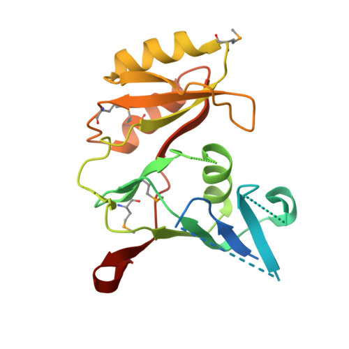
    Ribose-5-phosphate isomerase A (RpiA; EC 5.3.1.6) interconverts ribose-5-phosphate and ribulose-5-phosphate. This enzyme plays essential roles in carbohydrate anabolism and catabolism; it is ubiquitous and highly conserved. The structure of RpiA from Escherichia coli was solved by multiwavelength anomalous diffraction (MAD) phasing, and refined to 1.5 A resolution (R factor 22.4%, R(free) 23.7%). RpiA exhibits an alpha/beta/(alpha/beta)/beta/alpha fold, some portions of which are similar to proteins of the alcohol dehydrogenase family. The two subunits of the dimer in the asymmetric unit have different conformations, representing the opening/closing of a cleft. Active site residues were identified in the cleft using sequence conservation, as well as the structure of a complex with the inhibitor arabinose-5-phosphate at 1.25 A resolution. A mechanism for acid-base catalysis is proposed.


  • Organizational Affiliation

    Structural Biology Center, Argonne National Laboratory, 9700 South Cass Avenue, Building 202, Argonne, IL 60439, USA.


Macromolecules
Find similar proteins by:  (by identity cutoff)  |  3D Structure
Entity ID: 1
MoleculeChains Sequence LengthOrganismDetailsImage
RIBOSE 5-PHOSPHATE ISOMERASE
A, B
219Escherichia coliMutation(s): 0 
EC: 5.3.1.6
UniProt
Find proteins for P0A7Z0 (Escherichia coli (strain K12))
Explore P0A7Z0 
Go to UniProtKB:  P0A7Z0
Entity Groups  
Sequence Clusters30% Identity50% Identity70% Identity90% Identity95% Identity100% Identity
UniProt GroupP0A7Z0
Sequence Annotations
Expand
  • Reference Sequence
Small Molecules
Ligands 1 Unique
IDChains Name / Formula / InChI Key2D Diagram3D Interactions
ABF
Query on ABF

Download Ideal Coordinates CCD File 
C [auth A],
D [auth B]
5-O-phosphono-beta-D-arabinofuranose
C5 H11 O8 P
KTVPXOYAKDPRHY-SQOUGZDYSA-N
Modified Residues  1 Unique
IDChains TypeFormula2D DiagramParent
MSE
Query on MSE
A, B
L-PEPTIDE LINKINGC5 H11 N O2 SeMET
Binding Affinity Annotations 
IDSourceBinding Affinity
ABF Binding MOAD:  1O8B Ki: 2.10e+6 (nM) from 1 assay(s)
PDBBind:  1O8B Ki: 2.10e+6 (nM) from 1 assay(s)
Experimental Data & Validation

Experimental Data

  • Method: X-RAY DIFFRACTION
  • Resolution: 1.25 Å
  • R-Value Free: 0.240 
  • R-Value Work: 0.224 
  • R-Value Observed: 0.224 
  • Space Group: P 1
Unit Cell:
Length ( Å )Angle ( ˚ )
a = 42.049α = 90.23
b = 42.4β = 100.98
c = 60.195γ = 98.98
Software Package:
Software NamePurpose
REFMACrefinement
MOSFLMdata reduction
SCALAdata scaling
MOLREPphasing

Structure Validation

View Full Validation Report


Ligand Structure Quality Assessment 


Entry History 

Revision History  (Full details and data files)

  • Version 1.0: 2003-01-24
    Type: Initial release
  • Version 1.1: 2011-07-13
    Changes: Advisory, Version format compliance
  • Version 1.2: 2019-08-21
    Changes: Data collection, Database references, Derived calculations
  • Version 2.0: 2020-07-29
    Type: Remediation
    Reason: Carbohydrate remediation
    Changes: Atomic model, Data collection, Derived calculations, Other, Structure summary
  • Version 2.1: 2023-12-13
    Changes: Data collection, Database references, Refinement description, Structure summary