1NEU

STRUCTURE OF MYELIN MEMBRANE ADHESION MOLECULE P0


Experimental Data Snapshot

  • Method: X-RAY DIFFRACTION
  • Resolution: 1.90 Å
  • R-Value Free: 0.268 
  • R-Value Work: 0.215 
  • R-Value Observed: 0.215 

wwPDB Validation   3D Report Full Report


This is version 1.2 of the entry. See complete history


Literature

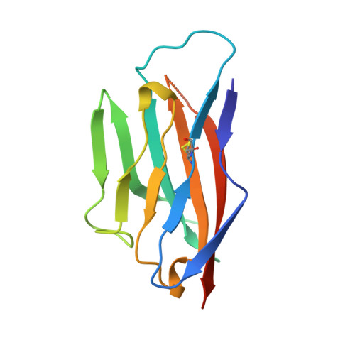
Crystal structure of the extracellular domain from P0, the major structural protein of peripheral nerve myelin.

Shapiro, L.Doyle, J.P.Hensley, P.Colman, D.R.Hendrickson, W.A.

(1996) Neuron 17: 435-449

  • DOI: https://doi.org/10.1016/s0896-6273(00)80176-2
  • Primary Citation of Related Structures:  
    1NEU

  • PubMed Abstract: 

    P0, the major protein of peripheral nerve myelin, mediates membrane adhesion in the spiral wraps of the myelin sheath. We have determined the crystal structure of the extracellular domain from P0 (P0ex) at 1.9 A resolution. P0ex is folded like a typical immunoglobulin variable-like domain; five residues at the C-terminus are disordered, suggesting a flexible linkage to the membrane. The requirements for crystallization of P0ex are similar to those for maintaining the native extracellular spacing of adjacent myelin lamellae; thus, given the self-adhesive character of P0ex, the crystal itself may reveal some of the natural interactions that occur between P0 molecules in myelin. The structure leads to the suggestion that P0 extracellular domains may emanate from the membrane surface as tetramers that link to tetramers on the opposing membrane surface, to result in the formation of networks of molecules. We report analytical ultracentrifugation data for P0ex that support this idea.


  • Organizational Affiliation

    Department of Biochemistry and Molecular Biophysics, Columbia University, New York, New York 10032, USA.


Macromolecules
Find similar proteins by:  (by identity cutoff)  |  3D Structure
Entity ID: 1
MoleculeChains Sequence LengthOrganismDetailsImage
MYELIN P0 PROTEIN124Rattus norvegicusMutation(s): 0 
Membrane Entity: Yes 
UniProt
Find proteins for P06907 (Rattus norvegicus)
Explore P06907 
Go to UniProtKB:  P06907
Entity Groups  
Sequence Clusters30% Identity50% Identity70% Identity90% Identity95% Identity100% Identity
UniProt GroupP06907
Sequence Annotations
Expand
  • Reference Sequence
Experimental Data & Validation

Experimental Data

  • Method: X-RAY DIFFRACTION
  • Resolution: 1.90 Å
  • R-Value Free: 0.268 
  • R-Value Work: 0.215 
  • R-Value Observed: 0.215 
  • Space Group: I 4 2 2
Unit Cell:
Length ( Å )Angle ( ˚ )
a = 88.9α = 90
b = 88.9β = 90
c = 91.6γ = 90
Software Package:
Software NamePurpose
X-PLORrefinement

Structure Validation

View Full Validation Report


Entry History 

Deposition Data

Revision History  (Full details and data files)

  • Version 1.0: 1997-05-15
    Type: Initial release
  • Version 1.1: 2008-03-24
    Changes: Version format compliance
  • Version 1.2: 2011-07-13
    Changes: Version format compliance