1N9E

Crystal structure of Pichia pastoris Lysyl Oxidase PPLO


Experimental Data Snapshot

  • Method: X-RAY DIFFRACTION
  • Resolution: 1.65 Å
  • R-Value Free: 0.187 
  • R-Value Work: 0.161 
  • R-Value Observed: 0.160 

wwPDB Validation   3D Report Full Report


Ligand Structure Quality Assessment 


This is version 2.1 of the entry. See complete history


Literature

The Crystal Structure of Pichia pastoris Lysyl Oxidase

Duff, A.P.Cohen, A.E.Ellis, P.J.Kuchar, J.A.Langley, D.B.Shepard, E.M.Dooley, D.M.Freeman, H.C.Guss, J.M.

(2003) Biochemistry 42: 15148-15157

  • DOI: https://doi.org/10.1021/bi035338v
  • Primary Citation of Related Structures:  
    1N9E

  • PubMed Abstract: 

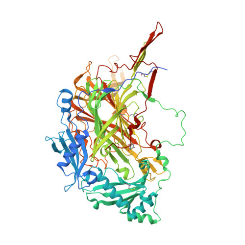
    Pichia pastoris lysyl oxidase (PPLO) is unique among the structurally characterized copper amine oxidases in being able to oxidize the side chain of lysine residues in polypeptides. Remarkably, the yeast PPLO is nearly as effective in oxidizing a mammalian tropoelastin substrate as is a true mammalian lysyl oxidase isolated from bovine aorta. Thus, PPLO is functionally related to the copper-containing lysyl oxidases despite the lack of any significant sequence similarity with these enzymes. The structure of PPLO has been determined at 1.65 A resolution. PPLO is a homodimer in which each subunit contains a Type II copper atom and a topaquinone cofactor (TPQ) formed by the posttranslational modification of a tyrosine residue. While PPLO has tertiary and quaternary topologies similar to those found in other quinone-containing copper amine oxidases, its active site is substantially more exposed and accessible. The structural elements that are responsible for the accessibility of the active site are identified and discussed.


  • Organizational Affiliation

    School of Molecular and Microbial Biosciences, University of Sydney, Sydney, NSW, Australia.


Macromolecules
Find similar proteins by:  (by identity cutoff)  |  3D Structure
Entity ID: 1
MoleculeChains Sequence LengthOrganismDetailsImage
LYSYL OXIDASE
A, B, C, D
787Komagataella pastorisMutation(s): 0 
EC: 1.4.3.13
UniProt
Find proteins for Q96X16 (Komagataella pastoris)
Explore Q96X16 
Go to UniProtKB:  Q96X16
Entity Groups  
Sequence Clusters30% Identity50% Identity70% Identity90% Identity95% Identity100% Identity
UniProt GroupQ96X16
Sequence Annotations
Expand
  • Reference Sequence
Oligosaccharides

Help

Entity ID: 2
MoleculeChains Length2D Diagram Glycosylation3D Interactions
2-acetamido-2-deoxy-beta-D-glucopyranose-(1-4)-2-acetamido-2-deoxy-beta-D-glucopyranose
E, F, G, H
2N-Glycosylation
Glycosylation Resources
GlyTouCan:  G42666HT
GlyCosmos:  G42666HT
GlyGen:  G42666HT
Small Molecules
Ligands 4 Unique
IDChains Name / Formula / InChI Key2D Diagram3D Interactions
NAG
Query on NAG

Download Ideal Coordinates CCD File 
AB [auth D]
BB [auth D]
I [auth A]
J [auth A]
K [auth A]
AB [auth D],
BB [auth D],
I [auth A],
J [auth A],
K [auth A],
KA [auth C],
L [auth A],
LA [auth C],
MA [auth C],
NA [auth C],
W [auth B],
X [auth B],
Y [auth B],
YA [auth D],
Z [auth B],
ZA [auth D]
2-acetamido-2-deoxy-beta-D-glucopyranose
C8 H15 N O6
OVRNDRQMDRJTHS-FMDGEEDCSA-N
SO4
Query on SO4

Download Ideal Coordinates CCD File 
DA [auth B]
EA [auth B]
FA [auth B]
FB [auth D]
GA [auth B]
DA [auth B],
EA [auth B],
FA [auth B],
FB [auth D],
GA [auth B],
GB [auth D],
HA [auth B],
HB [auth D],
IA [auth B],
IB [auth D],
JA [auth B],
JB [auth D],
KB [auth D],
LB [auth D],
P [auth A],
Q [auth A],
R [auth A],
RA [auth C],
S [auth A],
SA [auth C],
T [auth A],
TA [auth C],
U [auth A],
UA [auth C],
V [auth A],
VA [auth C],
WA [auth C],
XA [auth C]
SULFATE ION
O4 S
QAOWNCQODCNURD-UHFFFAOYSA-L
CU
Query on CU

Download Ideal Coordinates CCD File 
AA [auth B],
CB [auth D],
M [auth A],
OA [auth C]
COPPER (II) ION
Cu
JPVYNHNXODAKFH-UHFFFAOYSA-N
CA
Query on CA

Download Ideal Coordinates CCD File 
BA [auth B]
CA [auth B]
DB [auth D]
EB [auth D]
N [auth A]
BA [auth B],
CA [auth B],
DB [auth D],
EB [auth D],
N [auth A],
O [auth A],
PA [auth C],
QA [auth C]
CALCIUM ION
Ca
BHPQYMZQTOCNFJ-UHFFFAOYSA-N
Modified Residues  1 Unique
IDChains TypeFormula2D DiagramParent
TPQ
Query on TPQ
A, B, C, D
L-PEPTIDE LINKINGC9 H9 N O5TYR
Experimental Data & Validation

Experimental Data

  • Method: X-RAY DIFFRACTION
  • Resolution: 1.65 Å
  • R-Value Free: 0.187 
  • R-Value Work: 0.161 
  • R-Value Observed: 0.160 
  • Space Group: C 1 2 1
Unit Cell:
Length ( Å )Angle ( ˚ )
a = 248.442α = 90
b = 121.125β = 124.64
c = 151.841γ = 90
Software Package:
Software NamePurpose
MOSFLMdata reduction
SCALAdata scaling
AMoREphasing
REFMACrefinement
CCP4data scaling

Structure Validation

View Full Validation Report


Ligand Structure Quality Assessment 


Entry History 

Deposition Data

Revision History  (Full details and data files)

  • Version 1.0: 2004-01-13
    Type: Initial release
  • Version 1.1: 2008-04-28
    Changes: Version format compliance
  • Version 1.2: 2011-07-13
    Changes: Non-polymer description, Version format compliance
  • Version 1.3: 2014-10-29
    Changes: Derived calculations
  • Version 1.4: 2018-02-07
    Changes: Advisory, Experimental preparation
  • Version 2.0: 2020-07-29
    Type: Remediation
    Reason: Carbohydrate remediation
    Changes: Atomic model, Data collection, Derived calculations, Structure summary
  • Version 2.1: 2023-10-25
    Changes: Advisory, Data collection, Database references, Refinement description, Structure summary