Three-dimensional structure of rabbit liver [Cd7]metallothionein-2a in aqueous solution determined by nuclear magnetic resonance.
Arseniev, A., Schultze, P., Worgotter, E., Braun, W., Wagner, G., Vasak, M., Kagi, J.H., Wuthrich, K.(1988) J Mol Biol 201: 637-657
- PubMed: 3418714
- DOI: https://doi.org/10.1016/0022-2836(88)90644-4
- Primary Citation of Related Structures:
1MRB, 2MRB - PubMed Abstract:
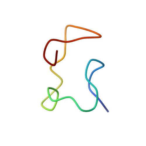
In previous work the metal-polypeptide co-ordinative bonds in the major protein species of a reconstituted [113Cd7]metallothionein-2 preparation from rabbit liver in aqueous solution were determined, the secondary polypeptide structure was found to contain several half-turns and 3(10)-helical segments, and a preliminary characterization of the overall polypeptide backbone fold in the beta-domain containing the three-metal cluster, and the alpha-domain containing the four-metal cluster, was obtained. Using a new, more extensive set of nuclear magnetic resonance data these earlier structures were improved by new structure calculations. The new experimental data consist of distance constraints from measurements of nuclear Overhauser effects, and dihedral angle constraints derived from both coupling constants and nuclear Overhauser effects. The structure calculations were performed with the program DISMAN. Since no information on the orientation of the two domains relative to each other could be obtained, the structure calculations were performed separately for the alpha-domain and the beta-domain. The average of the pairwise root-mean-square distances among the 20 structures with the least residual violations of input constraints was 2.9 A for the beta-domain and 1.4 A for the alpha-domain (1 A = 0.1 nm). The overall chirality of the polypeptide fold is right-handed for the beta-domain and left-handed for the alpha-domain. For each of the seven metal ions the local chirality of the co-ordination of the four cysteinyl Sy atoms is clearly defined. The improved structures of both domains show the previously noted differences relative to the recently published crystal structure of metallothionein-2a from rat liver.
Organizational Affiliation:
Institut für Molekularbiologie und Biophysik, Eidgenössische Technische Hochschule-Hönggerberg, Zürich, Switzerland.