1MN2

MANGANESE PEROXIDASE SUBSTRATE BINDING SITE MUTANT E35Q, D179N


Experimental Data Snapshot

  • Method: X-RAY DIFFRACTION
  • Resolution: 2.00 Å
  • R-Value Work: 0.187 
  • R-Value Observed: 0.187 

wwPDB Validation   3D Report Full Report


Ligand Structure Quality Assessment 


This is version 2.0 of the entry. See complete history


Literature

Crystal structures of substrate binding site mutants of manganese peroxidase.

Sundaramoorthy, M.Kishi, K.Gold, M.H.Poulos, T.L.

(1997) J Biol Chem 272: 17574-17580

  • DOI: https://doi.org/10.1074/jbc.272.28.17574
  • Primary Citation of Related Structures:  
    1MN1, 1MN2

  • PubMed Abstract: 

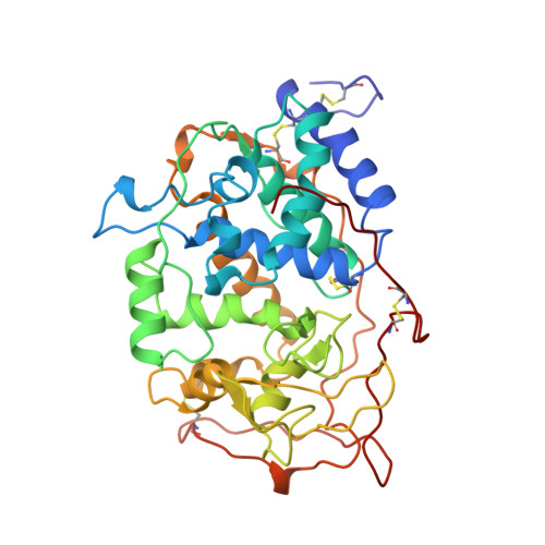
    Manganese peroxidase (MnP), an extracellular heme enzyme from the lignin-degrading basidiomycetous fungus, Phanerochaete chrysosporium, catalyzes the oxidation of MnII to MnIII. The latter, acting as a diffusible redox mediator, is capable of oxidizing a variety of lignin model compounds. The proposed MnII binding site of MnP consists of a heme propionate, three acidic ligands (Glu-35, Glu-39, and Asp-179), and two water molecules. Using crystallographic methods, this binding site was probed by altering the amount of MnII bound to the protein. Crystals grown in the absence of MnII, or in the presence of EDTA, exhibited diminished electron density at this site. Crystals grown in excess MnII exhibited increased electron density at the proposed binding site but nowhere else in the protein. This suggests that there is only one major MnII binding site in MnP. Crystal structures of a single mutant (D179N) and a double mutant (E35Q,D179N) at this site were determined. The mutant structures lack a cation at the MnII binding site. The structure of the MnII binding site is altered significantly in both mutants, resulting in increased access to the solvent and substrate.


  • Organizational Affiliation

    Department of Molecular Biology & Biochemistry and Physiology & Biophysics, University of California, Irvine, California 92697-3900, USA.


Macromolecules
Find similar proteins by:  (by identity cutoff)  |  3D Structure
Entity ID: 1
MoleculeChains Sequence LengthOrganismDetailsImage
MANGANESE PEROXIDASE357Phanerodontia chrysosporiumMutation(s): 0 
Gene Names: MNP1
EC: 1.11.1.13
UniProt
Find proteins for Q02567 (Phanerodontia chrysosporium)
Explore Q02567 
Go to UniProtKB:  Q02567
Entity Groups  
Sequence Clusters30% Identity50% Identity70% Identity90% Identity95% Identity100% Identity
UniProt GroupQ02567
Sequence Annotations
Expand
  • Reference Sequence
Oligosaccharides

Help

Entity ID: 2
MoleculeChains Length2D Diagram Glycosylation3D Interactions
2-acetamido-2-deoxy-beta-D-glucopyranose-(1-4)-2-acetamido-2-deoxy-beta-D-glucopyranose
B
2N-Glycosylation
Glycosylation Resources
GlyTouCan:  G42666HT
GlyCosmos:  G42666HT
GlyGen:  G42666HT
Experimental Data & Validation

Experimental Data

  • Method: X-RAY DIFFRACTION
  • Resolution: 2.00 Å
  • R-Value Work: 0.187 
  • R-Value Observed: 0.187 
  • Space Group: C 1 2 1
Unit Cell:
Length ( Å )Angle ( ˚ )
a = 163.24α = 90
b = 45.97β = 97.16
c = 53.57γ = 90
Software Package:
Software NamePurpose
XENGENdata collection
XENGENdata reduction
X-PLORmodel building
X-PLORrefinement
XENGENdata scaling
X-PLORphasing

Structure Validation

View Full Validation Report


Ligand Structure Quality Assessment 


Entry History 

Deposition Data

Revision History  (Full details and data files)

  • Version 1.0: 1997-09-04
    Type: Initial release
  • Version 1.1: 2008-03-24
    Changes: Version format compliance
  • Version 1.2: 2011-07-13
    Changes: Non-polymer description, Version format compliance
  • Version 1.3: 2018-03-07
    Changes: Data collection, Other
  • Version 2.0: 2020-07-29
    Type: Remediation
    Reason: Carbohydrate remediation
    Changes: Atomic model, Data collection, Database references, Derived calculations, Structure summary