Solution Structure of a Parallel Left-Handed Double-Helical Gramicidin-A Determined by 2D 1H NMR.
Chen, Y., Tucker, A., Wallace, B.A.(1996) J Mol Biol 264: 757
- PubMed: 8980684
- DOI: https://doi.org/10.1006/jmbi.1996.0675
- Primary Citation of Related Structures:
1MIC - PubMed Abstract:
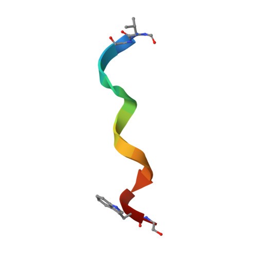
The structure of a parallel left-handed double-helical form of gramicidin was detected by circular dichroism spectroscopy and determined using 500 and 600 MHz NMR in CaCl2/methanol solution. Measurements of TOCSY, DQF-COSY and NOESY spectra were converted into 604 distance and 48 torsional angle constraints for structure calculations. Stereospecific assignments and chi 1 angles were calculated using 3J alpha beta, d alpha beta (i,i), dN beta(i,i) and dN gamma(i,i). chi 2 angles were determined using d alpha beta(i,i), dN beta(i,i), d beta delta(i,i), dN gamma(i,i) and d alpha gamma(i,i). The calculations of initial structures were performed using the distance geometry/simulated annealing method in XPLOR. The initial structures were further refined and energy minimized using simulated annealing/molecular dynamics methods. Back-calculations for every generated structure were also performed to check their consistency with the experimental data. 187 final structures with no violations above the threshold conditions (0.05 A, 5 degrees, 5 degrees, 0.5 A and 5 degrees for bonds, angles, improper, NOE and cdihe, respectively) were produced from the 200 initial structures. Twenty structures with the lowest NOE energies were used for further analysis. The average r.m.s. deviations for the 20 structures are 0.64 A for backbone and 1.1 A for all non-hydrogen atoms. Gramicidin in this form, with approximately 5.7 residues per turn, is a parallel double helical dimer. The length along the helix axis is about 30 A and the inner pore diameter varies from 1 to 2 A. It is different from all other gramicidin structures determined to date. The presence of Ca2+ stabilises a conformation that prevents the binding of monovalent cations. It is likely that this structure is related to a non-channel, antibiotic role of gramicidin.
Organizational Affiliation:
Department of Crystallography, Birkbeck College, University of London, UK.