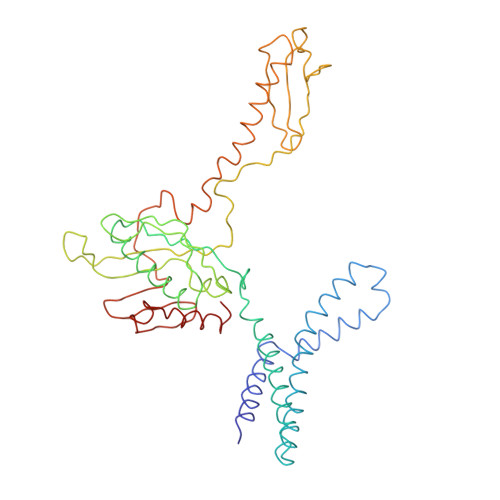
1MI6

Docking of the modified RF2 X-ray structure into the Low Resolution Cryo-EM map of RF2 E.coli 70S Ribosome


Experimental Data Snapshot

  • Method: ELECTRON MICROSCOPY
  • Resolution: 12.8 Å
  • Aggregation State: PARTICLE 
  • Reconstruction Method: SINGLE PARTICLE 

wwPDB Validation   3D Report Full Report


This is version 1.5 of the entry. See complete history


Literature

A cryo-electron microscopic study of ribosome-bound termination factor RF2

Rawat, U.B.S.Zavialov, A.V.Sengupta, J.Valle, M.Grassucci, R.A.Linde, J.Vestergaard, B.Ehrenberg, M.Frank, J.

(2003) Nature 421: 87-90

  • DOI: https://doi.org/10.1038/nature01224
  • Primary Citation of Related Structures:  
    1MI6, 1MVR

  • PubMed Abstract: 

    Protein synthesis takes place on the ribosome, where genetic information carried by messenger RNA is translated into a sequence of amino acids. This process is terminated when a stop codon moves into the ribosomal decoding centre (DC) and is recognized by a class-1 release factor (RF). RFs have a conserved GGQ amino-acid motif, which is crucial for peptide release and is believed to interact directly with the peptidyl-transferase centre (PTC) of the 50S ribosomal subunit. Another conserved motif of RFs (SPF in RF2) has been proposed to interact directly with stop codons in the DC of the 30S subunit. The distance between the DC and PTC is approximately 73 A. However, in the X-ray structure of RF2, SPF and GGQ are only 23 A apart, indicating that they cannot be at DC and PTC simultaneously. Here we show that RF2 is in an open conformation when bound to the ribosome, allowing GGQ to reach the PTC while still allowing SPF-stop-codon interaction. The results indicate new interpretations of accuracy in termination, and have implications for how the presence of a stop codon in the DC is signalled to PTC.


  • Organizational Affiliation

    Howard Hughes Medical Institute, Health Research, Inc., Empire State Plaza, Albany NY 12201-0509, USA.


Macromolecules
Find similar proteins by:  (by identity cutoff)  |  3D Structure
Entity ID: 1
MoleculeChains Sequence LengthOrganismDetailsImage
peptide chain release factor RF-2365Escherichia coli K-12Mutation(s): 0 
UniProt
Find proteins for P07012 (Escherichia coli (strain K12))
Explore P07012 
Go to UniProtKB:  P07012
Entity Groups  
Sequence Clusters30% Identity50% Identity70% Identity90% Identity95% Identity100% Identity
UniProt GroupP07012
Sequence Annotations
Expand
  • Reference Sequence
Experimental Data & Validation

Experimental Data

  • Method: ELECTRON MICROSCOPY
  • Resolution: 12.8 Å
  • Aggregation State: PARTICLE 
  • Reconstruction Method: SINGLE PARTICLE 
EM Software:
TaskSoftware PackageVersion
RECONSTRUCTIONSPIDER

Structure Validation

View Full Validation Report


Entry History 

Deposition Data

Revision History  (Full details and data files)

  • Version 1.0: 2003-01-14
    Type: Initial release
  • Version 1.1: 2008-04-28
    Changes: Version format compliance
  • Version 1.2: 2011-07-13
    Changes: Source and taxonomy, Version format compliance
  • Version 1.3: 2017-02-01
    Changes: Structure summary
  • Version 1.4: 2018-07-18
    Changes: Data collection
  • Version 1.5: 2024-02-14
    Changes: Data collection, Database references, Refinement description