1MHS

Model of Neurospora crassa proton ATPase


Experimental Data Snapshot

  • Method: ELECTRON CRYSTALLOGRAPHY
  • Resolution: 8.00 Å

wwPDB Validation   3D Report Full Report


This is version 1.4 of the entry. See complete history


Literature

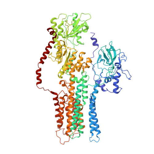
Structure, mechanism and regulation of the Neurospora plasma membrane H+-ATPase

Kuhlbrandt, W.Zeelen, J.Dietrich, J.

(2002) Science 297: 1692-1696

  • DOI: https://doi.org/10.1126/science.1072574
  • Primary Citation of Related Structures:  
    1MHS

  • PubMed Abstract: 

    Proton pumps in the plasma membrane of plants and yeasts maintain the intracellular pH and membrane potential. To gain insight into the molecular mechanisms of proton pumping, we built an atomic homology model of the proton pump based on the 2.6 angstrom x-ray structure of the related Ca2+ pump from rabbit sarcoplasmic reticulum. The model, when fitted to an 8 angstrom map of the Neurospora proton pump determined by electron microscopy, reveals the likely path of the proton through the membrane and shows that the nucleotide-binding domain rotates by approximately 70 degrees to deliver adenosine triphosphate (ATP) to the phosphorylation site. A synthetic peptide corresponding to the carboxyl-terminal regulatory domain stimulates ATPase activity, suggesting a mechanism for proton transport regulation.


  • Organizational Affiliation

    Max-Planck-Institut für Biophysik, Heinrich-Hoffmann-Str. 7, 60528 Frankfurt am Main, Germany.


Macromolecules
Find similar proteins by:  (by identity cutoff)  |  3D Structure
Entity ID: 1
MoleculeChains Sequence LengthOrganismDetailsImage
Plasma Membrane ATPase
A, B
920Neurospora crassaMutation(s): 0 
EC: 3.6.3.6
Membrane Entity: Yes 
UniProt
Find proteins for P07038 (Neurospora crassa (strain ATCC 24698 / 74-OR23-1A / CBS 708.71 / DSM 1257 / FGSC 987))
Explore P07038 
Go to UniProtKB:  P07038
Entity Groups  
Sequence Clusters30% Identity50% Identity70% Identity90% Identity95% Identity100% Identity
UniProt GroupP07038
Sequence Annotations
Expand
  • Reference Sequence
Experimental Data & Validation

Experimental Data

  • Method: ELECTRON CRYSTALLOGRAPHY
  • Resolution: 8.00 Å
  • Space Group: P 3 2 1
Unit Cell:
Length ( Å )Angle ( ˚ )
a = 167α = 90
b = 167β = 90
c = 250γ = 120
EM Software:
TaskSoftware PackageVersion
RECONSTRUCTIONMRC

Structure Validation

View Full Validation Report


Entry History 

Deposition Data

Revision History  (Full details and data files)

  • Version 1.0: 2002-09-18
    Type: Initial release
  • Version 1.1: 2008-04-28
    Changes: Version format compliance
  • Version 1.2: 2011-07-13
    Changes: Version format compliance
  • Version 1.3: 2018-07-18
    Changes: Author supporting evidence, Data collection
  • Version 1.4: 2024-02-14
    Changes: Data collection, Database references