1MC8

Crystal Structure of Flap Endonuclease-1 R42E mutant from Pyrococcus horikoshii


Experimental Data Snapshot

  • Method: X-RAY DIFFRACTION
  • Resolution: 3.10 Å
  • R-Value Free: 0.279 
  • R-Value Work: 0.190 
  • R-Value Observed: 0.197 

wwPDB Validation   3D Report Full Report


This is version 1.4 of the entry. See complete history


Literature

Molecular Structure and Novel DNA Binding Sites Located in Loops of Flap Endonuclease-1 from Pyrococcus horikoshii

Matsui, E.Musti, K.V.Abe, J.Yamazaki, K.Matsui, I.Harata, K.

(2002) J Biol Chem 277: 37840-37847

  • DOI: https://doi.org/10.1074/jbc.M205235200
  • Primary Citation of Related Structures:  
    1MC8

  • PubMed Abstract: 

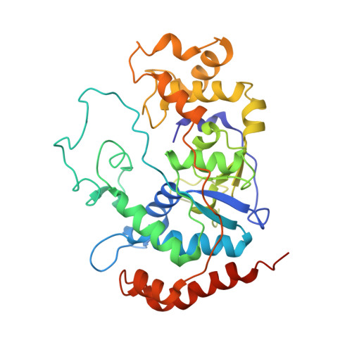
    The crystal structure of flap endonuclease-1 from Pyrococcus horikoshii (phFEN-1) was determined to a resolution of 3.1 A. The active cleft of the phFEN-1 molecule is formed with one large loop and four small loops. We examined the function of the conserved residues and positively charged clusters on these loops by kinetic analysis with 45 different mutants. Arg(40) and Arg(42) on small loop 1, a cluster Lys(193)-Lys(195) on small loop 2, and two sites, Arg(94) and Arg(118)-Lys(119), on the large loop were identified as binding sites. Lys(87) on the large loop may play significant roles in catalytic reaction. Furthermore, we successfully elucidated the function of the four DNA binding sites that form productive ES complexes specific for each endo- or exo-type hydrolysis, probably by bending the substrates. For the endo-activity, Arg(94) and Lys(193)-Lys(195) located at the top and bottom of the molecule were key determinants. For the exo-activity, all four sites were needed, but Arg(118)-Lys(119) was dominant. The major binding sites for both the nick substrate and double-stranded DNA might be the same.


  • Organizational Affiliation

    Biological Information Research Center and the Gene Discovery Research Center, National Institute of Advanced Industrial Science and Technology, Higashi 1-1-1, Tsukuba, Ibaraki 305-566, Japan.


Macromolecules
Find similar proteins by:  (by identity cutoff)  |  3D Structure
Entity ID: 1
MoleculeChains Sequence LengthOrganismDetailsImage
Flap Endonuclease-1
A, B
343Pyrococcus horikoshiiMutation(s): 1 
UniProt
Find proteins for O50123 (Pyrococcus horikoshii (strain ATCC 700860 / DSM 12428 / JCM 9974 / NBRC 100139 / OT-3))
Explore O50123 
Go to UniProtKB:  O50123
Entity Groups  
Sequence Clusters30% Identity50% Identity70% Identity90% Identity95% Identity100% Identity
UniProt GroupO50123
Sequence Annotations
Expand
  • Reference Sequence
Experimental Data & Validation

Experimental Data

  • Method: X-RAY DIFFRACTION
  • Resolution: 3.10 Å
  • R-Value Free: 0.279 
  • R-Value Work: 0.190 
  • R-Value Observed: 0.197 
  • Space Group: P 31
Unit Cell:
Length ( Å )Angle ( ˚ )
a = 62.67α = 90
b = 62.67β = 90
c = 180.69γ = 120
Software Package:
Software NamePurpose
MOSFLMdata reduction
TRUNCATEdata reduction
X-PLORmodel building
X-PLORrefinement
CCP4data scaling
X-PLORphasing

Structure Validation

View Full Validation Report


Entry History 

Deposition Data

Revision History  (Full details and data files)

  • Version 1.0: 2002-10-16
    Type: Initial release
  • Version 1.1: 2008-04-28
    Changes: Version format compliance
  • Version 1.2: 2011-07-13
    Changes: Version format compliance
  • Version 1.3: 2021-11-10
    Changes: Database references
  • Version 1.4: 2023-10-25
    Changes: Data collection, Refinement description