1MC5

Ternary complex of Human glutathione-dependent formaldehyde dehydrogenase with S-(hydroxymethyl)glutathione and NADH


Experimental Data Snapshot

  • Method: X-RAY DIFFRACTION
  • Resolution: 2.60 Å
  • R-Value Free: 0.249 
  • R-Value Work: 0.197 
  • R-Value Observed: 0.197 

wwPDB Validation   3D Report Full Report


Ligand Structure Quality Assessment 


This is version 1.4 of the entry. See complete history


Literature

Human glutathione-dependent formaldehyde dehydrogenase. Structural changes associated with Ternary Complex formation

Sanghani, P.C.Bosron, W.F.Hurley, T.D.

(2002) Biochemistry 41: 15189-15194

  • DOI: https://doi.org/10.1021/bi026705q
  • Primary Citation of Related Structures:  
    1MC5

  • PubMed Abstract: 

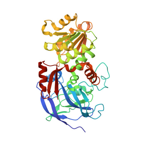
    Human glutathione-dependent formaldehyde dehydrogenase plays an important role in the metabolism of glutathione adducts such as S-(hydroxymethyl)glutathione and S-nitrosoglutathione. The role of specific active site residues in binding these physiologically important substrates and the structural changes during the catalytic cycle of glutathione-dependent formaldehyde dehydrogenase was examined by determining the crystal structure of a ternary complex with S-(hydroxymethyl)glutathione and the reduced coenzyme to 2.6 A resolution. The formation of the ternary complex caused the movement of the catalytic domain toward the coenzyme-binding domain. This represents the first observation of domain closure in glutathione-dependent formaldehyde dehydrogenase in response to substrate binding. A water molecule adjacent to the 2'-ribose hydroxyl of NADH suggests that the alcohol proton is relayed to solvent directly from the coenzyme, rather than through the action of the terminal histidine residue as observed in the proton relay system for class I alcohol dehydrogenases. S-(Hydroxymethyl)glutathione is directly coordinated to the active site zinc and forms interactions with the highly conserved residues Arg114, Asp55, Glu57, and Thr46. The active site zinc has a tetrahedral coordination environment with Cys44, His66, and Cys173 as the three protein ligands in addition to S-(hydroxymethyl)glutathione. This is in contrast to zinc coordination in the binary coenzyme complex where all of the ligands were contributed by the enzyme and included Glu67 as the fourth protein ligand. This change in zinc coordination is accomplished by an approximately 2.3 A movement of the catalytic zinc.


  • Organizational Affiliation

    Center for Structural Biology, Department of Biochemistry and Molecular Biology, Indiana University School of Medicine, Indianapolis, Indiana 46202, USA.


Macromolecules
Find similar proteins by:  (by identity cutoff)  |  3D Structure
Entity ID: 1
MoleculeChains Sequence LengthOrganismDetailsImage
Alcohol dehydrogenase class III chi chain
A, B
374Homo sapiensMutation(s): 0 
Gene Names: ADH5
EC: 1.1.1.1 (PDB Primary Data), 1.2.1.1 (PDB Primary Data)
UniProt & NIH Common Fund Data Resources
Find proteins for P11766 (Homo sapiens)
Explore P11766 
Go to UniProtKB:  P11766
PHAROS:  P11766
GTEx:  ENSG00000197894 
Entity Groups  
Sequence Clusters30% Identity50% Identity70% Identity90% Identity95% Identity100% Identity
UniProt GroupP11766
Sequence Annotations
Expand
  • Reference Sequence
Small Molecules
Ligands 5 Unique
IDChains Name / Formula / InChI Key2D Diagram3D Interactions
NAD
Query on NAD

Download Ideal Coordinates CCD File 
H [auth A],
M [auth B]
NICOTINAMIDE-ADENINE-DINUCLEOTIDE
C21 H27 N7 O14 P2
BAWFJGJZGIEFAR-NNYOXOHSSA-N
AHE
Query on AHE

Download Ideal Coordinates CCD File 
I [auth A]2-AMINO-4-[1-CARBOXYMETHYL-CARBAMOYL)-2-HYDROXYMETHYLSULFANYL-ETHYLCARBAMOYL]-BUTYRIC ACID
C11 H19 N3 O7 S
PIUSLWSYOYFRFR-BQBZGAKWSA-N
PO4
Query on PO4

Download Ideal Coordinates CCD File 
F [auth A],
G [auth A],
L [auth B]
PHOSPHATE ION
O4 P
NBIIXXVUZAFLBC-UHFFFAOYSA-K
ZN
Query on ZN

Download Ideal Coordinates CCD File 
C [auth A],
D [auth A],
J [auth B],
K [auth B]
ZINC ION
Zn
PTFCDOFLOPIGGS-UHFFFAOYSA-N
K
Query on K

Download Ideal Coordinates CCD File 
E [auth A]POTASSIUM ION
K
NPYPAHLBTDXSSS-UHFFFAOYSA-N
Experimental Data & Validation

Experimental Data

  • Method: X-RAY DIFFRACTION
  • Resolution: 2.60 Å
  • R-Value Free: 0.249 
  • R-Value Work: 0.197 
  • R-Value Observed: 0.197 
  • Space Group: P 43 21 2
Unit Cell:
Length ( Å )Angle ( ˚ )
a = 78.656α = 90
b = 78.656β = 90
c = 311.428γ = 90
Software Package:
Software NamePurpose
DENZOdata reduction
SCALEPACKdata scaling
CNSrefinement
CNSphasing

Structure Validation

View Full Validation Report


Ligand Structure Quality Assessment 


Entry History 

Deposition Data

Revision History  (Full details and data files)

  • Version 1.0: 2003-03-04
    Type: Initial release
  • Version 1.1: 2008-04-28
    Changes: Version format compliance
  • Version 1.2: 2011-07-13
    Changes: Version format compliance
  • Version 1.3: 2018-04-04
    Changes: Data collection
  • Version 1.4: 2024-02-14
    Changes: Data collection, Database references, Derived calculations, Refinement description