NMR analysis of the monomeric form of a mutant unliganded bovine neurophysin: comparison with the crystal structure of a neurophysin dimer.
Nguyen, T.L., Breslow, E.(2002) Biochemistry 41: 5920-5930
- PubMed: 11980496
- DOI: https://doi.org/10.1021/bi012067k
- Primary Citation of Related Structures:
1L5C, 1L5D - PubMed Abstract:
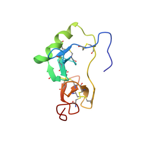
Determination of the structure of the unliganded monomeric state of neurophysin is central to an understanding of the allosteric relationship between neurophysin peptide-binding and dimerization. We examined this state by NMR, using the weakly dimerizing H80E mutant of bovine neurophysin-I. The derived structure, to which more than one conformer appeared to contribute, was compared with the crystal structure of the unliganded des 1-6 bovine neurophysin-II dimer. Significant conformational differences between the two proteins were evident in the orientation of the 3,10 helix, in the 50-58 loop, in beta-turns, and in specific intrachain contacts between amino- and carboxyl domains. However, both had similar secondary structures, in independent confirmation of earlier circular dichroism studies. Previously suggested interactions between the amino terminus and the 50-58 loop in the monomer were also confirmed. Comparison of the observed differences between the two proteins with demonstrated effects of dimerization on the NMR spectrum of bovine neurophysin-I, and preliminary investigation of the effects of dimerization on H80E spectra, allowed tentative distinction between the contributions of sequence and self-association differences to the difference in conformation. Regions altered by dimerization encompass most binding site residues, providing a potential explanation of differences in binding affinity between the unliganded monomeric and dimeric states. Differences between monomer and dimer states in turns, interdomain contacts, and within the interdomain segment of the 50-58 loop suggest that the effects of dimerization on intrasubunit conformation reflect the need to adjust the relative positions of the interface segments of the two domains for optimal interaction with the adjacent subunit and/or reflect the dual role of some residues as participants both at the interface and in interdomain contacts.
Organizational Affiliation:
Department of Biochemistry, Weill Medical College of Cornell University, 1300 York Avenue, New York, New York 10021, USA.