Crystal structure of the Bse634I restriction endonuclease: comparison of two enzymes recognizing the same DNA sequence.
Grazulis, S., Deibert, M., Rimseliene, R., Skirgaila, R., Sasnauskas, G., Lagunavicius, A., Repin, V., Urbanke, C., Huber, R., Siksnys, V.(2002) Nucleic Acids Res 30: 876-885
- PubMed: 11842098
- DOI: https://doi.org/10.1093/nar/30.4.876
- Primary Citation of Related Structures:
1KNV - PubMed Abstract:
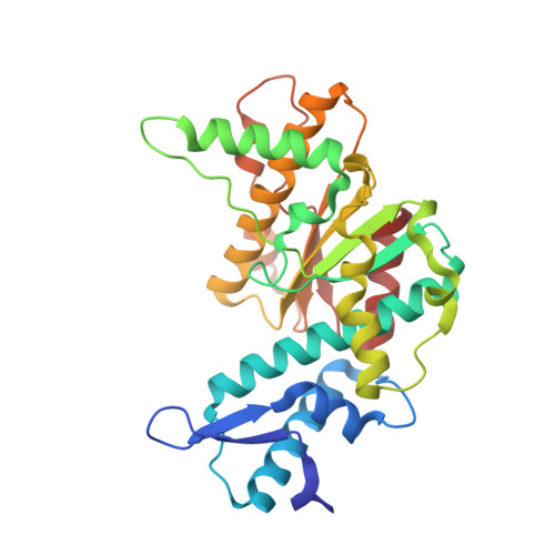
Crystal structures of Type II restriction endonucleases demonstrate a conserved common core and active site residues but diverse structural elements involved in DNA sequence discrimination. Comparative structural analysis of restriction enzymes recognizing the same nucleotide sequence might therefore contribute to our understanding of the structural diversity of specificity determinants within restriction enzymes. We have solved the crystal structure of the Bacillus stearothermophilus restriction endonuclease Bse634I by the multiple isomorphous replacement technique to 2.17 A resolution. Bse634I is an isoschisomer of the Cfr10I restriction enzyme whose crystal structure has been reported previously. Comparative structural analysis of the first pair of isoschisomeric enzymes revealed conserved structural determinants of sequence recognition and catalysis. However, conformations of the N-terminal subdomains differed between Bse634I/Cfr10I, suggesting a rigid body movement that might couple DNA recognition and catalysis. Structural similarities extend to the quaternary structure level: crystal contacts suggest that Bse634I similarly to Cfr10I is arranged as a tetramer. Kinetic analysis reveals that Bse634I is able to interact simultaneously with two recognition sites supporting the tetrameric architecture of the protein. Thus, restriction enzymes Bse634I, Cfr10I and NgoMIV, recognizing overlapping nucleotide sequences, exhibit a conserved tetrameric architecture that is of functional importance.
Organizational Affiliation:
Max-Planck Institut für Biochemie, Abt. Strukturforschung, Am Klopferspitz 18a, D-82152 Martinsried (bei München), Germany. grazulis@ibt.lt